Download
1 / 12
180 likes | 961 Views
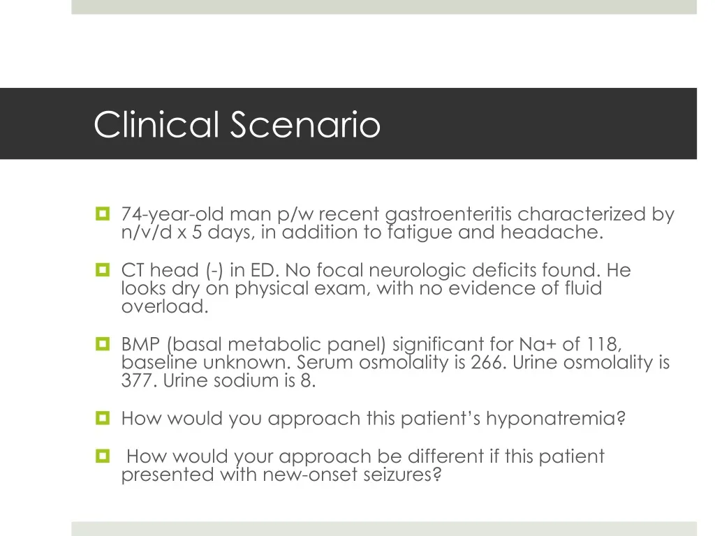
Clinical Scenario. 74-year-old man p/w recent gastroenteritis characterized by n/v/d x 5 days, in addition to fatigue and headache. CT head (-) in ED. No focal neurologic deficits found. He looks dry on physical exam, with no evidence of fluid overload.

E N D
Clinical Scenario • 74-year-old man p/w recent gastroenteritis characterized by n/v/d x 5 days, in addition to fatigue and headache. • CT head (-) in ED. No focal neurologic deficits found. He looks dry on physical exam, with no evidence of fluid overload. • BMP (basal metabolic panel) significant for Na+ of 118, baseline unknown. Serum osmolality is 266. Urine osmolality is 377. Urine sodium is 8. • How would you approach this patient’s hyponatremia? • How would your approach be different if this patient presented with new-onset seizures?
Lecture Objectives • Hyponatremia • Clinical manifestations • Diagnostic approach • Clinical Scenario discussed
Hyponatremia Defined • Definition: Serum Na+ <135 meq/L • Generally associated with decreased osmolality to <275 • Most common electrolyte abnormality. • Caused by retention of water • Usually a drop in osmolality will suppress ADH to allow excretion of the excess water via dilute urine • Most forms of hyponatremia are associated with elevated ADH (whether appropriate or inappropriate), which concentrates urine
Signs & Symptoms • More profound when the decrease in sodium is very large or occurs rapidly(i.e. over hours) • Generally asymptomatic if Na+ level >125, postural hypotension only symptom in early phases and neurological symptoms occur when Na+ level >110 to 115. • Symptoms include: • Headache • Nausea, vomiting • Muscle cramps • Disorientation, depressed reflexes, lethargy, restlessness • Seizure, coma, permanent brain damage, respiratory arrest, brainstem herniation & death • Serious complications are more commonly seen after surgery, and in menstruating women
Symptoms • Tachycardia • Postural hypotension • Dry mucous membranes • Soft sunken eyeballs • Decreased skin turgor • Decreased urine output
Approach to Hyponatremia • 1st assess volume status • Is the patient volume overloaded, depleted, or euvolemic? • 2nd assess osmolality (hyper, iso, or hypo) • Is the blood concentrated? For hypotonic hyponatremia, continue to 3rd step: • 3rdassess urinary sodium excretion • Is the urine concentrated? *Remember VOU – volume status, osmolality, and urine studies
STEP 1 – (V) Volume Status • 1st assess volume status (extracellular fluid volume) • hyponatremiahas 3 main etiologies: • Hypovolemic – both H2O and Na decreased (H20 < Na) • Consider obvious losses from diarrhea, vomiting, dehydration, malnutrition, etc • Euvolemic – H20 increased and Na stable • Consider siADH, thyroid disease, primary polydipsia • Hypervolemic – H20 increased and Na increased (H2O > Na) • Consider obvious CHF, cirrhosis, renal failure
STEP 2 - (O) Osmolality • 2nd assess osmolality hyper, iso, or hypo • Hypotonic hyponatremia = warrants further workup, especially when there is no obvious fluid overload or depletion • Serum Osmolality: lab value or calculation – in mosm/kg • =(2 x Na+) + (glucose/18) + (BUN/2.8) + (ethanol)/4.6 • Hypertonic - >295 • hyperglycemia, mannitol, glycerol • Isotonic- 280-295 • pseudo-hyponatremia from elevated lipids or protein • Hypotonic - <280 • excess fluid intake, low solute intake, renal disease, siADH, hypothyroidism, adrenal insufficiency, CHF, cirrhosis, etc.
STEP 3 – (U) Urine Studies • For euvolemic hyponatremia, check urine osmolality • Urine osmolality <100 - excess water intake • Primary polydipsia • Urine osmolality >100 - impaired renal concentration • siADH, hypothyroidism, cortisol deficiency • Check urine sodium & calculate FeNa % ( percentage of the sodium filtered by the kidney which is excreted in the urine) • A low urine sodium (<10) and low FeNa (<1%) implies the kidneys are appropriately reabsorbing sodium • A high urine sodium (>20) and high FeNa (>1%) implies the kidneys are not functioning properly
Treatment of Hyponatremia • Be CAUTIOUS with correction: • 0.5 meq/L increase per every hour initially • Do not increase Na more than 10 meq/L in 24 hrs or 18 meq/L in 48 hrs • Treatment varies greatly by etiology of hyponatremia.
Clinical Scenario - Conclusion • 74-year-old man p/w recent gastroenteritis characterized by n/v/d x 5 days, in addition to fatigue and headache. • BMP significant for Na+ of 118, baseline unknown. Serum osmolality is 266. Urine osmolality is 377. • How would you approach this patient’s hyponatremia? The steps: • 1) Serum osmolality – 266, decreased (hypotonic) • 2) Urine osmolality –377, increased (>100) • 3) Volume status – hypovolemic • 4) Treatment: Mild symptoms, correct slowly w/ isotonic saline • How would your approach be different if this patient presented with new-onset seizures? • For symptomatic, severe hyponatremia, more rapid correction using 3% normal saline
Case study • 42 y old male 2 days history of vomiting diarrhoea. Intake was only water. Weak unable to stand, with pulse 114/min, BP was 100/55, BMP was as follows: • Sodium 131 • K 3 • Urea 17.8 (2.5- 8) • Creatinine 150 (40 -130)