Download
1 / 50
630 likes | 1.33k Views
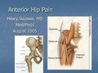
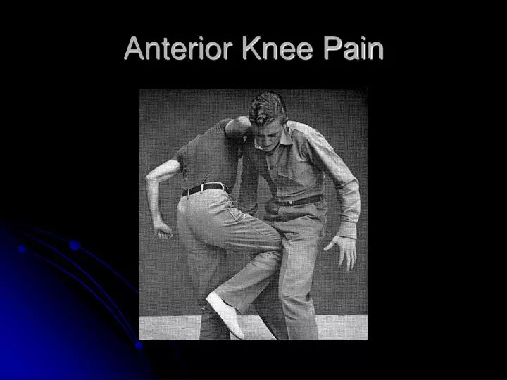
Anterior Knee Pain. Patellofemoral pain syndrome Trauma-Dislocation Osteoarthrosis Cartilage abnormalities Osteochondritis dissecans Bipartite patella-Dorsal defect of the patella. Synovial Plica Extensor mechanism tears Bursitis Osgood –Schlatter Disease.

E N D
Patellofemoral pain syndrome Trauma-Dislocation Osteoarthrosis Cartilage abnormalities Osteochondritis dissecans Bipartite patella-Dorsal defect of the patella Synovial Plica Extensor mechanism tears Bursitis Osgood –Schlatter Disease. Excessive lateral pressure syndrome Anterior Knee Pain
Objectives • Discuss basic anatomy and biomechanics of the patellofemoral joint • Understand imaging methods and limitations of these imaging methods used to assess the patellofemoral joint. • Be familiar with basic terminology and measurements used to describe the patellofemoral joint in order to communicate with the clinicians accurately and effectively. • Have a working differential diagnosis of anterior knee pain
History • Skeletal findings prove that the knee joint has been in existence for over 320 million years • The Eryops, the ancestors of the reptiles, birds and mammals, seems to be the first creature in the animal kingdom with a bicondylar knee joint. • The patellofemoral joint, however, only began to develop some 65 million years ago.
Facets • The posterior surface of the patella articulates with the trochlear groove along the anterior surface of the femoral condyles to form the patellofemoral joint. • The posterior patella has a medial and lateral facet. A variable, usually small, odd facet lies along the medial border of the patella.
Wrisberg Variants • Type 1 patellae have concave medial and lateral facets approximately equal in size (10%) • Type 2 also have concave facets, but the medial facet is smaller than the lateral (65%) • Type 3 have a small convex medial facet (25%)
Passive Stabilizers • The patellar ligament and the medial and lateral patellar retinacula form the passive stabilizers of the patella. • The retinacula have deep and superficial layers and can have a bilaminar appearance. • The retinacula provide significant stabilizing support to the patella.
Passive Stabilizers • On the medial side, the medial patellofemoral ligament has been shown to be the major passive restraint preventing lateral patellar dislocation • The medial patellofemoral ligament arises between the adductor tubercle (the insertion of the adductor magnus tendon), and the medial epicondyle (the site of origin of the tibial collateral ligament). • The ligament then runs forward just deep to the distal vastus medialis obliquus muscle to attach to the superior two thirds of the medial patella margin. Adductor tendon Vastus Medialis Obliquus MPFL Superficial Medial collateral ligament
Medial Patellofemoral Ligament • (a) image taken immediately inferior to the adductor tubercle demonstrates a normal femoral origin of the MPFL (open arrow). The distal vastus medialis obliquus muscle (arrowhead) lies anteriorly. • (b) image just inferior to (a) demonstrates the proximal origin of the tibial collateral ligament (open arrowhead). Note that the medial patellar retinaculum (open arrow) can have a normal bilaminar appearance. Gradient Echo
Dynamic Stabilizers • The four quadriceps muscles form the active stabilizers of the patella. • The inferior portions of the vastus medialis and lateralis muscles form small muscle groups with a distinct oblique orientation of their fibers, the vastus medialis obliquus and the vastus lateralis obliquus muscles.
Biomechanics • The patella is the largest sesamoid bone • By displacing the fulcrum of motion of the extensor mechanism anterior to the femur, the patellofemoral articulation produces a mechanical advantage increasing the force of the quadriceps muscles in extending the knee. • The patella also centralizes the divergent forces of the quadriceps muscle and transmits the tension around the femur to the patellar tendon.
Biomechanics • Considerable force is transmitted across the patellofemoral joint • The force varies from half body weight during walking, up to 25 times body weight on lifting a weight with the knees flexed at 90°
Biomechanics • In the fully extended knee the patella lies superior to the trochlear cartilage. • As the knee flexes to 30°, the patella begins to engage with the trochlea. • Between 30 and 90° of flexion, first the inferior and then the superior patella cartilage articulates with the trochlear cartilage. • Beyond 120°, contact is reduced between the patella and trochlea.
Q Angle • The Q angle is formed between a line joining the anterior superior iliac spine and the center of the patella, and a line joining the center of the patella and the tibial tuberosity. • Normal angle 10-12 degrees in males and 15-18 in females • Questionable validity
Techniques for performing the axial radiograph of the patella • The prone technique (a) requires knee flexion >90°, and therefore eliminates subluxation in most patients with tracking abnormality. • Supine techniques are more valuable for assessment of patella alignment and include those of Laurin et al. (b) with the knee flexed at 20°, and Merchant et al. (c) with the knee flexed at 45°. The Merchant technique may be performed with the beam direction reversed (d), which eliminates the need for a special cassette holder. • To perform a weight-bearing axial view (e) a specially designed knee support is required, but this may provide a more physiologic assessment, of patellofemoral alignment
Sulcus Angle • Used to measure trochlear depth • A line is drawn from the lowest point of the intercondylar sulcus, B, to the highest points of the lateral and medial femoral condyles, A and C. The angle between lines AB and BC is the sulcus angle. • Normal range 126–150°
Congruence Angle • Used to measure lateral patellar displacement • To measure the congruence angle (curved arrow) in (a), the sulcus angle is bisected to produce a reference line, and the angle is measured between this reference and a line joining the apex of the sulcus, B, and the lowest point of the patellar articular surface, D. • In the normal knee, point D should lie no more than 16° lateral to the bisected sulcus angle.
Lateral Patellar Displacement • (b) Measured by drawing a line joining the summits of the medial and lateral femoral condyles and dropping a perpendicular to this at the level of the summit of the medial condyle. The distance of the medial margin of the patella from this perpendicular is measured (arrowheads). • In the normal knee the medial patellar margin should lie no more than 1 mm lateral to the perpendicular.
Lateral Patellofemoral Angle • Used to measure patellar tilt. • (c) (curved arrow) is the angle between a line joining the apices of the femoral condyles and a line joining the limits of the lateral patellar facet. The angle is taken to be normal when it opens laterally, and abnormal when it opens medially.
Patellofemoral Measurements on the Lateral Radiograph • In grade I alignment (normal) the median ridge of the patella (open arrow) lies posterior to the lateral facet (curved arrow). On a lateral radiograph the median ridge and lateral facet form two separate borders which appear slightly concave. • With mild patellar tilt (grade II) the median ridge and lateral facet line up on the lateral views so that only one border is seen. • With further tilt (grade III), the lateral facet projects posterior to the median ridge and appears convex. • Normal lateral radiograph of the knee. The depth of the trochlear groove may be measured 1 cm distal to its upper limit (arrows). Less than 5 mm is considered dysplastic.
Patellar Height • For the Insall-Salvati method the patellar ligament length is divided by the maximal diagonal length of the patella on the lateral radiograph.The ratio here is 1.5 (>1.2 indicates patella alta). • (b) A modified index, which is less sensitive to variation in patella morphology, is calculated as the distance between the inferior articular surface of the patella and the patellar ligament insertion divided by the length of the patella articular surface. The ratio is measured at 2.2 (>2 indicates patella alta).
Axial Evaluation • The right knee shows no subluxation. • The left knee shows osteochondral irregularity to the medial patella with a small separated adjacent bony fragment (arrowhead) as well as an osteochondral fragment at the lateral femoral condyle (arrow), all consistent with prior patellar dislocation.
Patellofemoral pain syndrome Trauma-Dislocation Osteoarthrosis Cartilage abnormalities Osteochondritis dissecans Bipartite patella-Dorsal defect of the patella Synovial Plica Extensor mechanism tears Bursitis Osgood –Schlatter Disease. Excessive lateral pressure syndrome Anterior Knee Pain
Patellofemoral Pain Syndrome • Loosly used term to describe anterior knee pain that is thought to be due to malalignment and maltracking issues. • Symptoms include anterior knee pain and giving way.
Definitions • Patellofemoral alignment refers to the static relationship between the patella and the trochlea at a given degree of knee flexion. • Patellofemoral tracking refers to the dynamic patellofemoral alignment during knee motion.
Patellofemoral Pain Syndrome • Most common diagnosis in outpatients presenting with knee pain • 16-25 % of injuries in runners • 11% of musculoskeletal complaints in the office
Patellofemoral Pain Syndrome • Current perspective is that this is a clinical diagnosis and imaging studies are not necessary before starting treatment. • Radiography is recommended in patients with a history of trauma or surgery, those with an effusion, those older than 50 years of age, and those that do not improve with treatment.
Limitations of Radiology • Clear definitions of maltracking are limited by the fact that clinical and radiologic measures described are often abnormal in asymptomatic knees and within described normal ranges in symptomatic knees. • Measures of alignment will vary depending on the degree of knee flexion. • Imaging studies of the patellofemoral joint for tracking should focus on the first 30-45 degrees of flexion. In early flexion is when anatomical factors such as patella alta, trochlear dysplasia and abnormalities of the soft tissue restraints of the patella have the most pronounced effect in producing abnormal tracking.
Patellofemoral pain syndrome Trauma-Dislocation Osteoarthrosis Cartilage abnormalities Osteochondritis dissecans Bipartite patella-Dorsal defect of the patella Synovial Plica Extensor mechanism tears Bursitis Osgood –Schlatter Disease. Excessive lateral pressure syndrome Anterior Knee Pain
Lateral Patellar Dislocation • Anteroposterior radiograph of the knee showing a laterally dislocated patella. The patella usually spontaneously reduces and this appearance is rare. • The patella is reduced, but note the osteochondral fragment adjacent to the medial patella and the small concave defect at the medial patellar margin.
Lateral Patellar Dislocation • Three weeks after acute transient lateral patellar dislocation demonstrates a concave impaction deformity (small white arrows) of the medial patella. • There is a contusion (asterisk) at the lateral femoral condyle. Note the complete tear (open white arrow) at the patellar insertion of the medial patellar retinaculum.
Lateral Patellar Dislocation Courtesy of T. Dog Hughes
Medial Patellofemoral Ligament • (a) image taken immediately inferior to the adductor tubercle demonstrates a normal femoral origin of the MPFL (open arrow). The distal vastus medialis obliquus muscle (arrowhead) lies anteriorly. • (b) image just inferior to (a) demonstrates the proximal origin of the medial collateral ligament (open arrowhead). Note that the medial patellar retinaculum (open arrow) shows a bilaminar appearance. Gradient Echo
Lateral Patellar Dislocation • Image of the knee 4 days after acute transient lateral patellar dislocation. There is complete disruption of the medial patellofemoral ligament from its femoral attachment (thin white arrow). • Note the concave impaction deformity of the inferomedial patella (black arrow) with marrow contusion. Axial FS T2
Lateral Patellar Dislocation Courtesy of T. Dog Hughes
Lateral Patellar Dislocation • Image 3 weeks after acute transient lateral patellar dislocation demonstrates edema surrounding the distal vastus medialis obliquus muscle T2
Osteochondritis Dissecans • There is focal full-thickness cartilage loss, as well as loss of a fragment of subchondral bone, as evidenced by loss of the black stripe representing the subchondral bone plate within the lesion. • Deep to the lesion there is edema.
Dorsal Defect of Patella Courtesy of T. Dog Hughes
Dorsal Defect of the Patella • Defect in the subchondral bone of the superior patella. • Note that the overlying cartilage is thickened over the defect to produce a near normal articular surface T1 T2
Bipartite Patella • Accessory ossification center at the superolateral patella. • Axial fat-saturated T2-weighted image demonstrates that the overlying cartilage appears intact.
Excessive Lateral Pressure Syndrome • There is marked lateral patellar tilt but little subluxation and there is full-thickness cartilage loss and marrow edema confined to the lateral patella facet. Note the normal cartilage thickness at the medial patella (white arrows).
Conclusion • Discuss basic anatomy and biomechanics of the patellofemoral joint • Understand imaging methods and limitations of these imaging methods used to assess the patellofemoral joint. • Be familiar with basic terminology and measurments used to describe the patellofemoral joint in order to communicate with the clinicians acurately and effectively. • Have a working differential diagnosis of anterior knee pain
Bibliography • Conway W, Hayes C, Loughran T, et al. Cross-sectional Imaging of the Patellofemoral Joint and Surrounding Structures. Radiographics 1991; 11:195-217. • Techlenburg K, Dejour D, Hoser C, Fink C. Bony and cartilagintous anatomy of the patellofemoral joint. Knee Surg Sports Traumatol Arthrosc 2006; 14:235-240. • Shellock F, Mink J, Fox J. Patellofemoral Joint: Kinematic MR Imaging to Assess Tracking Abnormalities. Radiology 1988; 168:551-553 • Murray T, Dupont J, Fulkerson J. Axial and Lateral Radiographs in Evaluating Patellofemoral Malalignment. Amer J of Sports Medicine 1996; 27:580-584
Bibliography • Kujala U, Osterman K, Kormano M et al. Patellofemoral Relationships in Recurrent Patellar Dislocation. J bone Joint Surg 1989; 71:788-92 • Katchburian M, Bull A, Yi-Fen S, et al. Measurement of Patellar Tracking: Assessment and Analysis of the Literature. Clin Ortho Rel Res 2002; 412: 241-59. • MacIntyre, N, Hill N, Ellis R, et al. Patellofemoral Joint Kinematics in Indiividuals with and without Patellofemoral Pain Sydroms. J Bone Joint Surg 2006;88:2596-2605 • Dixit S, Difiori J, Burton M, et al. Management of Patellofemoral Pain sydrome. Am Fam Phys 2007;75:194-202.