Download
1 / 27
270 likes | 586 Views
Systemic Lupus Erythematosis. Patient. 14 year old girl Presented 2 mths previously with dyspnoea and chest pain at a regional hospital Initially treated as pneumonia Progressed to ICU admission 5 seizures in ward (started on phenytoin) TB as a child

E N D
Patient • 14 year old girl • Presented 2 mths previously with dyspnoea and chest pain at a regional hospital • Initially treated as pneumonia • Progressed to ICU admission • 5 seizures in ward (started on phenytoin) • TB as a child • No history of medicines, allergies, family risk • Development: Grade 6 – doing well
History continued • Transferred to tertiary hospital • Blood tests done, started on prednisone • Referred to Renal Clinic for renal biopsy
Results from clinic • ANA, ANF, Anti-Sm, Anti dsDNA, Ro, La Positive • Low C3, C4
On Examination • Weight: 44,7 kg (25th) • Height: 170 cm (75th) • Apyrexial, RR 18, HR 119, BP 96/70 • Marfanoid habitus, rash on face and body • Not anaemic, no oedema, shotty cervical glands • CVS: tachycardic • Resp, Abdo, neuro, ENT: normal • Musculo-skeletal: Marfanoid. No arthritis • Tanner stage 4
Urine • 4+ haemoglobin, no protein • Spun: scanty red cells and casts • Lab: 18 000 leukocytes < 1000 RBC’s Epithelial cells Gram negative bacilli
Laboratory investigations • Hb 11 (MCV 90.6), Plt 369 000 • WCC 5.6 (N61%, M4%, L35%) • U+E, CMP: normal • ESR 36, CRP 2.7 • INR, PTT, CK, Cholesterol, WR: normal
Special investigations • CXR: - residual opacification (RUL + RML) with fluid in fissure - ?some scoliosis - Heart size + aortic knuckle normal • ECG: normal • Echo: pending • Ophthalmology, dermatology consultations: pending
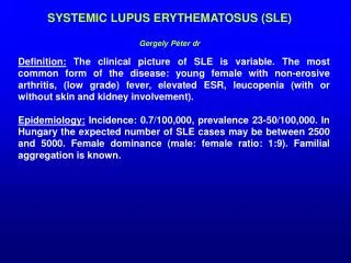
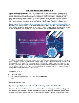
Introduction to SLE • Auto-immune disorder • Multisystem microvascular inflammation • Formation of autoantibodies • Chronic with relapsing and remitting course
Pathophysiology • Proposed mechanism for autoantibodies: • Defect in apoptosis • ↑cell death → disturbance in immune tolerance • Plasma + nuclear antigens displayed on cell surface • Dysregulated lymphocytes target Ag (normally intracellular) • Immune complexes form in microvasculature → complement activation + inflammation • Ag-Ab complexes deposit in basement membranes of skin and kidneys
Etiology • Unknown • At least 10 gene loci known to ↑ risk • Genetic predisposition (10x more in monozygotic twins) • Human leukocyte Ag: ↑ HLA-DR2, HLA-DR3 + HLA-B8 • Null complement alleles + congenital ↓complement (esp C4, C2 etc)
Epidemiology • Prevalence: 4 – 250/100 000 • Incidence US: 1/10 000 • Onset: before 8 yrs unusual • Female predominance (prepubertal 4:1, postpubertal 8:1) • Rare in black African population
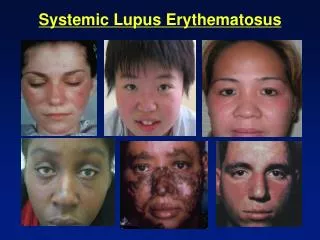
History • Constitutional – fatigue, fever, weight loss • Skin – malar rash, photosensitive, discoid lupus, alopecia, Raynaud phenomenon, livido reticularis • Musculoskeletal – arthralgia, myalgia, arthritis • Renal • Neuropsychiatric – headache, mood disorders, cognitive disorders, psychosis, seizures, TIA/ stroke, movement disorders, mononeuritis • Pulmonary – chest pain, dyspnoea • Gastrointestinal – Abdominal pain, jaundice • Cardiac – heart failure/chest pain • Haematological – multiple ‘cytopenias’ • Other – miscarriages, family history of autoimmune disease
On examination • Constitutional – lymphadenopathy, hepatosplenomegaly • Musculoskeletal – Jaccoud arthropathy • Dermatologic - capillaroscopy • Renal • Neuropsychiatric • Cardiopulmonary – friction rubs, pulmonary embolism, Libman-Sacks endocarditis • GIT – peritonitis, pancreatitis, mesenteric vasculitis
Diagnostic criteria American College of Rheumatology 4/11 criteria (sens 85%, specif 95%) “SOAP BRAIN MD” • Serositis – heart, lung, peritoneum • Oral ulcers – painless esp palate • Arthritis – non-erosive • Photosensitivity
Diagnostic criteria continued • Blood disorders - ↓RBC (Coombs +), PLT, WCC, Lymphocytes • Renal involvement – proteinuria /± casts • ANA – titer > 1:160 • Immunologic phenomena – LE cells, anti-dsDNA Ab, anti-Sm Ab, antiphospholipid Ab, false WR + • Neurological disorders – seizures/ psychosis • Malar rash – cheeks + nasal bridge • Discoid rash – rimmed with scaling, follicular plugging
Laboratory studies High clinical suspicion/ high ANA titres SLE Screen: • FBC and diff • S-creatinine • Urinalysis with microscopy • Basic inflammatory markers • Antibodies to dsDNA • Complement • ANA subtypes (anti-Sm, Ro, La, RNP Ab’s)
Autoantibody tests used in SLE • ANA – screening test (95% sensitivity) • Anti-dsDNA (high specifcity, sens 70%) • Anti-Sm (most specific Ab for SLE, 30% sens) • Anti-Ro/anti-La (15% in SLE patients, neonatal disease) • Anti-ribosomal P (uncommon, assoc lupus cerebritis) • Anti-RNP (overlap) • Anticardiolipin (antiphospholipid Ab syndrome) • Lupus Anticoagulant (antiphospholipid Ab syndrome) • Coombs test (Ab on RBC’s) • Anti-histone (drug-induced lupus)
Radiological studies • Joint x-rays: no erosions, periarticular osteopenia + soft tissue swelling • CXR/CT chest: interstitial lung disease, pneumonitis, pulmonary emboli, alveolar hemorrhage • CTBrain or Brain MRI ± angiography: lupus white matter changes, vasculitis or stroke • Echo: pericardial effusion, pulmonary hypertension or Libman-Sacks endocarditis
Invasive procedures • LP – nonspecific ↑cells + protein, ↓ glucose • Renal biopsy – prognosis and Rx • Skin biopsy
Differential diagnosis • Drug induced lupus erythematosis • Vasculitis • Leukemia • HIV • Multiple sclerosis • Parvovirus or other viral infections
Treatment principles • Depends on disease severity • Fever, skin, musculoskeletal and serositis = milder disease • CNS and renal involvement – aggressive Rx • Emergencies: - severe CNS involvement - systemic vasculitis - profound thrombocytopenia (TTP-like syndrome) - rapidly progressive nephritis - diffuse alveolar hemorrhage
Medications used • NSAIDS • Chloroquine • Steroids • Cyclophosphamide • Azathioprine • Mycophenolate • (Rituximab) • Plasma exchange/ IVIG
Preventive care • Medication-related (steroid) complications (Ca, vit D, bisphosphonates) • Aggressive BP and lipid control • Immunization (complement deficient) • Stress-dose steroid protocols for patients on maintenance corticosteroids (surgery/ infection) • Avoid UV exposure • Avoid estrogen therapies • Avoid sulfa-containing medications • Pregnancy planning
Prognosis • Benign to rapidly progressive • Better for isolated skin + musculoskeletal disease vs renal and CNS • Death rate 3X age-comparable general populationMortality • Nephritis (most within 5 yrs of symptoms) • Infectious (active SLE + Rx – most common) • CVS disease (50X more MI than other woman) • Malignancy (chronic inflammation + Rx)
Summary • Autoimmune disorder • Multiple manifestations • Aggressive investigation and treatment • Continued surveillance