Download
1 / 87
870 likes | 876 Views
Anatomy Ch. 12. The Lymphatic System and Body Defenses. The lymphatic system assists with the processes of the cardiovascular and immune systems. The lymphatic system consists of 2 parts A network of lymphatic vessels Transport fluids back to the blood that has escaped Lymphoid tissue

E N D
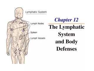
Anatomy Ch. 12 The Lymphatic System and Body Defenses
The lymphatic system assists with the processes of the cardiovascular and immune systems. • The lymphatic system consists of 2 parts • A network of lymphatic vessels • Transport fluids back to the blood that has escaped • Lymphoid tissue • Houses phagocytic cells and lymphocytes which play roles in body defense and resistance to disease.
Lymphatic Vessels • Fluid is forced out at the arterial end of capillary beds and most is reabsorbed at the venous end. • Fluid that remains behind in tissue spaces becomes part of the interstitial fluid. • This fluid must be carried back to the blood for there to be sufficient blood volume. • When excess fluid accumulates in the tissue swelling or edema occurs.
The function of the lymphatic vessels is to pick up the excess fluid and return it to the blood. • The fluid in the lymphatic vessels is called lymph. • The lymphatic vessels form a one way system and lymph flows only toward the heart.
Lymphatic capillaries are found between the tissue cells and blood capillaries. • The function of these capillaries is to absorb the leaked fluid. • Lymphatic capillaries have small valves that allow fluid to flow in one direction. • These valves work in a similar way to the valves of the heart and veins by preventing backflow. • The purpose of the valves is to prevent leaking and to force fluid in one direction along the vessel.
Large particles such as cell debris, bacteria, and viruses enter lymphatic capillaries easily. • The problem with this is that these foreign substances could use the lymph vessels to travel throughout the body. • This problem is resolved by the fact that lymph takes detours through lymph nodes where it is cleansed and examined by immune cells.
Lymph is transported from capillaries through successively larger vessels called collecting vessels. • Lymph is returned to blood in the venous system through 1 of 2 large ducts found in the thoracic region • Right lymphatic duct: right arm and right side of the head and thorax • Thoracic duct: rest of the body • Both ducts empty into the subclavian vein
Lymphatic vessels are like veins in that they… • Have thin walls • Have valves • Are under low pressure • Need the assistance of skeletal muscles and pressure changes • Smooth muscles in the walls of larger vessels also help to move lymph along
Lymph Nodes • Lymph nodes help protect the body by removing foreign material from the lymphatic system and by producing lymphocytes • Lymph is filtered through lymph nodes that are found along lymphatic vessels • Large clusters of nodes: • inguinal (groin) • axillary (armpit) • cervical (neck)
Cells found in lymph nodes: • Macrophages: engulf and destroy foreign particles • Lymphocytes: (WBCs) respond to foreign substances • The swelling of lymph nodes during an infection is a result of the trapping function of the nodes
Lymph nodes are less than an inch long • Each node is surrounded by a capsule • Trabeculae are strands that extend inward from the capsule. These strands divide the node into compartments. • The population of lymphocytes in the node continually changes • Lymphocytes form in red bone marrow and then migrate to the lymph nodes where they multiply rapidly.
The cortex is the outer part of the node • The cortex contains collections of lymphocytes called follicles • The follicles contain germinal centers • Germinal centers contain lymphocytes called B cells which produce plasma cells which release antibodies.
The rest of the cells in the cortex are lymphocytes called T cells • The T cells continuously circulate between the blood, lymph nodes, and lymphatic stream • The medulla of the node is the central region and contains macrophages.
Lymph enters the convex side of a node through afferent lymphatic vessels • The lymph then flows through a number of sinuses that meander through the node • Lymph exits at an area called the hilum through efferent lymphatic vessels
The flow of lymph through a node is slow because there are fewer efferent vessels than afferent. • This slow flow allows for the lymphocytes and macrophages to perform their functions.
Other Lymphatic Organs • Spleen • Filters and cleanses blood of bacteria and viruses • Contains lymphocytes • Most important function is to destroy worn out RBCs and return some of the products to the liver
Thymus • Functions at peak levels only during youth • Produces hormones that allow lymphocytes to mature. • Mature lymphocytes can distinguish between our cells and foreign substances • Tonsils • Trap and remove any bacteria or other foreign substances that enter the throat
Peyer’s patches • Found in the wall of the small intestine • Contains macrophages that capture and destroy bacteria in the digestive tract • Appendix • Offshoot of the large intestine • Function is similar to peyer’s patches
The peyer’s patches, appendix, and tonsils are part of a collection of lymphatic tissue called MALT • MALT stands for Mucosa Associated Lymphoid Tissue • MALT works to protect the upper respiratory and digestive tracts.
The Immune System • Innate defense system • Nonspecific defense system • Responds immediately to protect the body from all foreign substances • Functions in prevention • Includes: • 1st line – skin & mucous membranes • 2nd line – cells and chemicals
Adaptive Defense System • Specific defense system • Attacks particular foreign substances • Includes: • Lymphocytes • Macrophages • Dendritic cells • Must be primed by an initial exposure to a foreign substance before it can protect the body. • What this system lacks in speed it makes up for in precision.
The immune system protects us both directly and indirectly. • It protects us directly by cell attack • It protects us indirectly by releasing chemicals and antibodies • The resistance to disease is called immunity.
Innate Body Defenses • Surface membrane barriers • The body’s first line of defense is the skin and mucous membranes. • The skin is a strong physical barrier • The mucous membranes line the digestive, urinary, respiratory, and reproductive tracts • Some of the protective secretions include sebum, HCl acid, lysozyme, and mucus.
Cells and Chemicals (Internal Defenses) • The second line of defense • phagocytes • natural killer cells • inflammatory response • antimicrobial proteins (complement & interferon) • fever
Phagocytosis • Phagocytes engulf foreign particles • Cytoplasmic extensions bind to the particle and pull it inside of a vacuole • The vacuole is fused with a lysosome and the contents are digested. • Examples include macrophages and neutrophils.
Natural Killer Cells • Natural killer cells are a unique group of lymphocytes that can lyse and kill foreign cells. • Natural killer cells can react spontaneously on any target by recognizing certain sugars on the surface of foreign cells • They attack the cell membrane of foreign particles by releasing chemicals which cause the foreign cell to disintegrate.
Inflammatory Response • Triggered whenever body tissues are injured • The 4 most common signs of inflammation: • Redness • Heat • Swelling • Pain • When cells are injured they release chemicals which… • Cause blood vessels to dilate and capillaries to become leaky • Activate pain receptors • Attract phagocytes and WBCs to the area
The inflammatory response • Prevents the spread of damaging agents • Disposes of cell debris and pathogens • Sets the stage for repair • 3 steps of the inflammatory response • Neutrophils enter the blood • The neutrophils squeeze through the capillary wall • The neutrophils gather in the site of the injury and within an hour they are devouring any foreign material.
Monocytes follow the neutrophils and replace them in the process of disposal of cell debris. • Clotting proteins wall off the damaged area to prevent the spread of pathogens • Local heat increases metabolic rate and speeds up the defensive actions and repair process
Antimicrobial Proteins (complement and interferon) • Complement • Group of 20 plasma proteins produced by the liver. • Enhances the immune system. • Occurs when an antibody and antigen interact. • A group of 9 complement proteins (C1 – C9) undergo a series of reactions. • These reactions form membrane attack complexes (MACs). • Some proteins directly attack the pathogen. • MACs can cause 3 responses: • Lesions can form on the pathogen • Amplifying the inflammatory response • Opsonization (labeling for removal) by phagocytosis
Interferon • When a cell becomes infected by a virus it cannot save itself but it can help defend other cells that have not been infected. • Infected cells do this by secreting proteins called interferons • Interferon molecules diffuse to nearby cells and bind to their membranes • This binding causes the production of proteins that interfere with the ability of viruses to multiply in these healthy cells.
Fever • Abnormally high body temperature • Body temperature can rise in response to chemicals secreted by WBCs and macrophages that are exposed to foreign substances in the body • Mild or moderate fever benefits the body • Bacteria require large amounts of iron and zinc to multiply • During a fever the liver and spleen gather up these nutrients making them less available • Fever also increases metabolic rate which speeds up the repair process
Adaptive Defenses • Specific defenses • 3rd line of defense • 3 aspects • Antigen specific • Systemic – not restricted in infection site • Memory – recognizes and mounts a stronger attack
2 divisions • Humoral – antibody mediated • Cellular – cell mediated by lymphocytes • Direct cellular – lyse the foreign cell • Indirect cellular – releasing chemicals that enhance the response or activate other cells
Antigens • Substance capable of causing an immune response • Foreign intruders or nonself • Self antigens do not trigger an immune response
Cells of the Adaptive Defense System • Lymphocytes • B cells • produce antibodies • oversee humoral immunity • T cells • do not produce antibodies • Cell mediated immunity • Antigen Presenting Cells (APC) • Do not respond to specific antigens but help the lymphocytes that do
Lymphocytes • Produced in the red bone marrow • Whether a lymphocyte matures into a B or T cell depends on where in the body it becomes immunocompetent • T cells come from lymphocytes that mature in the thymus • Only the T cells that mature enough to identify foreign antigens survive. • Mature T cells must develop a self tolerance to normal body cells.
B cells mature in the bone marrow • Once a lymphocyte is immunocompetent it will be able to react to only 1 type of antigen • Our genes determine what foreign substances our immune system will be able to recognize and resist. • After T and B cells become immunocompetent they migrate to the lymph nodes and spleen where the antigen encounters will occur.
Antigen Presenting Cells (APC) • Major role is to engulf antigens and present fragments of them on their own surfaces where they can be recognized by T cells. • The major APCs are macrophages and dendritic cells • When macrophages present antigens, they activate T cells. • Activated T cells release chemicals that activate the macrophages making them true killers. • Macrophages tend to remain in lymphoid organs but T cells circulate throughout the body