Download
1 / 25
250 likes | 355 Views
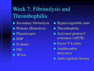
Hyperhomocyst(e)inemia and Thrombophilia. A major point of consensus was that no causal role of hyperhomocyst( e)inemia in venous or arterial thrombosis is not yet established. H omocysteine is a non–protein-forming sulfhydryl amino acid

E N D
A major point of consensus was that no causal role of hyperhomocyst( • e)inemia in venous or arterial thrombosis is not yet established
Homocysteine is a non–protein-forming sulfhydryl amino acid • Dietary methionine Homocysteine (Intracellular demethylation) • Homocysteine methionine (remethylation) • is derived from the reduction of • 5,10-methylene-tetrahydrofolate Methyltetrahydrofolate (MTHFR). • Methionine excess homocysteine may enter the transsulfuration pathway.
Hyperhomocystinemia :marked increase in atherothrombotic CVD and VTE. • 25% : vascular occlusive event by age 16 year & 50% by age 29 years. • Of these events, 32% are CVA, 4% are MI , 11% are peripheral arterial • occlusions, and 51% are VTEs. • The event rate is reduced by vitamin therapy in B6-responsive individuals.
The gene for MTHFR is located on chromosomal region 1p36. • Thermolabile mutant. • 12% of the population in the United States is homozygous • Estrogen-containing medications result in lower plasma homocysteine levels
New Concepts inCongenital Thrombophilia Galila Zaher (November 1999)
Congenital thrombophilia Inherited thrombophilia can be defined as a genetically determined tendency to venous thromboembolism. Genetic risk factors are now identified in 30- 50% of affected individuals.
Table 1. Risk factors for inherited thrombophilia: prevalence and relative risk for venous theombosis
Hyperhomocystinaemia Homocysteine is a sulphur containing A A. It is derived from methionine EAA. Homocysteine metabolism: Re methylation pathway MTHFR Trans sulphuration pathway C s Trans methylation pathway
Hyperhomocystinaemia Interest in homocysteine as a risk factor for vascular disease came from the early observations that such diseases are common in cases of classical homocystinuria.
Hyperhomocystinaemia Neural tube defects (NTPs) Arterial vascular disease Venous thromboembolism
HyperhomocystinaemiaNTDs. Neuronal tube defects occur in 1/1000 birth in USA. It has a complex trait interacting with environmental factors. Homozygousity for TL-MTHFR has been clearly shown to be a risk factor for spina bifida in 12% of the cases. Pre conceptional supplementation of folic acid could prevent up to 70% of NDTs. Folic acid 4 mg reduces the recurrence of NTDs (MRC vit study research gp) (Czei Zel, N Eng] Med 1992)
HyperhomocystinaemiaArterial vascular disease Potential mechanisms Oxidative damage to endothelial cells. Enhanced plt adhesion to endothelial cells Enhanced plt aggregation Plt accumulation and the formation of plt rich thrombous Inhibition of TM expression Increased procoagulant activity and reduced natural Anticoagulant (Wetch. N. Eng] Med 1998)
HyperhomocystinaemiaArterial vascular disease Metanalysis of published studies revealed that elevation in total plasma homocysteine were found to be an independent risk factor for all forms of arterial vascular disease. (Perry Advances in Haem. 1999)
HyperhomocystenaemiaArterial vascular disease * 15-40% of patients with conorary, cerebral of peripheral arterial disease have high plasma level of homocysteine > 20 moL/L. * The odd ratio for IHD is 1.4 for every 5 moL/L more than homocysteine median fasting adult males. * C677T mutation is a major cause of mild hyperhomocystinenia, but the mutation per se does not increase cvs disease risk
Hyperhomocystinaemiavenouss thromboembolism There is accumulating data to suggest that hyperhomocystinemia is a risk factor for VTE that is independent of coexisting abnormalities of the naturally occurring Anticoagulant. Homozygosity TL-MTHFR together with low folate level confers moderate risk factor. Hyperhomocystinaemia defined as plasma level >95% of control (18 M mol IL) was found in 16% of cases.
HyperhomcystinaemiaLaboratory evaluation Intra individual variability Inter Laboratory variability ELISA HPLC EIA
HyperhomocystinaemiaLaboratory evaluation There is an urgent need to improve analytical impression and decrease the difference among methods. An ideal homocysteine reference range based on targeting subject with highest serum Folate level is preferable to the population base range.
HyperhomocystinaemiaLaboratory evaluation Fasting total plasma homocysteine level Methionin loading dose. Serum Folate and B12 Pevel PCR C677T
HyperhomocystinaemiaLowering vit dose Plasma ThCY response to folic acid and Pyridoxin hydrochlorid The optimal homocysteine lowering vitamin dose and target ThCY are currently unknown. Proposed does. Folic acid 500-650 g Pyridoxin 100 mg B 12 0.4 mg.
Issues to be addressed What is the minimum dose of folic acid to prevent NTDs? Would grain fortification adapted in USA reduce NTDs, protects against arterial vascular disease? What is (are) the best test (s) to evalaute hyper homocystinemia?
Concluding Remarks • Folate supplementation reduces the occurance and recurrence of NTDs. • Elevated fasting plasma homocysteine is an independent risk factor for all forms of arterial vascular disease.
Concluding Remarks(Continue) • Homozygonosity for TL-MTHFR is not a significant risk factor for VTE per se • There is currently no strong argument to include MTHFR C677T genotyping during routine thrombophilia screen. • Combination of hypercystinaemia and FvL abnormality significantly increase the risk for VTE.