Download
1 / 5
50 likes | 200 Views
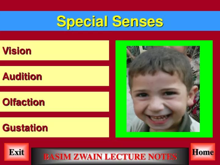
Special Senses. Vision. Audition. Olfaction. Gustation. Home. Exit. BASIM ZWAIN LECTURE NOTES. Vision. Image Formation. Background. Anatomy of the Eye. 3. Emmetropia (normal vision) a. Parallel light rays are focused on the retina without accommodation. Hyperopia (farsightedness)

E N D
Special Senses Vision Audition Olfaction Gustation Home Exit BASIM ZWAIN LECTURE NOTES
Vision Image Formation Background Anatomy of the Eye 3. Emmetropia (normal vision) a. Parallel light rays are focused on the retina without accommodation Hyperopia (farsightedness) a. Eye ball is too short b. Image is focused at a point behind the retina c. Lens can accommodate for distant objects but not for near d. Condition can be corrected with a convex lens (e.g., increase refractive power) Myopia (nearsightedness) a. Eye ball is too long b. Light rays converge in front of the retina c. Lens can accommodate for near objects but not distant d. Condition can be corrected with a concave lens Refraction by cornea Pupillary light reflex Additional terms and concepts Opthalmoscopic appearance Accommodation by the lens Cross sectional anatomy Gross anatomy 1. Distant objects a. Light rays run in parallel 2. Light rays slow a. Cornea b. Aqueous humor 3. Light rays bend a. Perpendicular to the angle (radius of the cornea) between the curve of the cornea and the plane they are traveling on Processes 1. Light energy is transduced into neural activity 2. Neural activity is processed by the brain Note: By way of analogy, you can imagine taking a picture with a camera. The eye is the camera, the retina, which is a specialized part of the brain at the back of the eye, is the film, and the parts of the brain that process visual information is the photoshop. 4. Focal distance a. Distance between the refractive surface and where the light rays converge b. Depends on the curvature of the cornea i. 2.4 cm ii. Distance between the cornea and the retina c. Vitreous humor--more viscous than the aqueous humor i. Lies between the lens and the retina ii. Provides spherical shape d. Retina i. Inner most layer of cells at the back of the eye ii. Transduces light energy into neural activity a.Optic disk (blind spot, no vision is possible) i. Blood vessels originate here. The vessels shadow the retina ii. Optic nerve fibers exit here iii. No photoreceptors b. Macula--area of the retina responsible for central vision (vs. peripheral) c. Fovea--center of the retina (where most of the cones are) 1. Pupil contribute to optical qualities of the eye a. Adjusts for different light levels b. Contributes to simultaneous focusing on near and distant objects 2. Accommodation alters light rays that would otherwise run in parallel a. Light rays are no longer focused on the retina by the cornea Sequence of events 1. Light entering the eye is focused on the retina 2. Retina converts light energy into neuronal activity 3. Axons of the retinal neurons are bundled to form the optic nerves 4. Visual information is distributed to several brain structures that perform different functions • Human visual systems permit light reflected off • distant objects to be: • Localized relative to the individual within his or • her environment • 2. Identified based on size, shape, color, and past • experience • 3. Perceived to be moving (or not) • 4. Detected in a wide variety of lighting conditions 3. Closing the aperture of the pupil a. Only light rays that are primarily in the center of the cornea and lens are allowed in b. These are generally not focused c. Permits seeing things in the foreground and background in focus e. Extraocular muscles--attached to the eye and skull and allow movement f. Conjunctiva--membrane inside the eyelid attached to the sclera g. Optic nerve--axons of the retina leaving the eye h. Cornea--transparent surface covering the iris and pupil 3. Contraction of ciliary muscles Tension on the suspensory ligaments is released b. Lens becomes rounded c. Greater the curvature provides greater the refraction Retina as seen through the pupil (aside: in photographs, the red appearance of the eye is actually the retina photographed. Double flash camera causes the pupil to constrict) • 1. External features of the eye • Pupil--opening that allows light to reach • the retina • b. Iris--circular muscle that controls • the diameter of the pupil • c. Aqueous humor--fluid behind the cornea • d. Sclera--outermost layer that forms the • eyeball 1. Visual field a. Total space that can be viewed by the retina i. 150 degrees ii. 90 on temporal side iii. 60 on the nasal side 2. Image formed on the back of the retina is reversed and inverted 1. Objects within 9 meters a. Light rays do not travel in parallel i. Some diverge 2. Lens adds refractive power Provided by changing the shape of the lens Processes 1. Refraction by the cornea 2. Accommodation by the lens 3. Pupillary light reflex a. Lens--transparent surface that contributes to the formation of images. b. Ciliary muscles--change the shape of the lens and allow focusing Structural levels 1. Gross anatomy 2. Opthalmoscopic appearance 3. Cross-section anatomy Home Exit BASIM ZWAIN LECTURE NOTES
Vision Microscopic Anatomy of the Retina 1. Rods--long, cylindrical, many disks a. Photopigment is in the disk b. Rods have a much higher pigment concentration c. 1000x more sensitive to light than cones d. Function in scotopic conditions i. Nighttime lighting e. All rods have the same pigment i. Rhodopsin • Specialized cells • of the retina convert • light energy into • neural activity • B. Cellular architecture • Of the retina C. Photoreceptors--two kinds based on appearance and function Characteristics: a. Photoreceptors are the only cells that respond to light b. Ganglion cells are the only output cells c. Light travels through the other cell layers to reach the photoreceptors d. At the back of the eye is a pigmented epithelium that absorbs any light not absorbed by the photoreceptors 5. Connectivity a. Central retina i. 1:1 (approximately) correspondence between photoreceptor and ganglion b. Peripheral retina i. Many photoreceptors (rods) converge on a single output ganglion cell c. Peripheral retina is more sensitive to light 2. Cones--shorter, tapering outer segment, relatively few disks a. Photopic conditions i. Daytime lighting ii. Primarily cones b. 3 different types of cones based on type of photopigment i. Pigments are differentially sensitive to wavelength of light Layers (3 primary, but there are subdivisions) a. Ganglion cell layer--cell bodies of the ganglion cells b. Inner nuclear layer--cell bodies of the bipolar cells c. Outer nuclear layer--cell bodies of the photoreceptors • 4. Distribution of rods and conesa • Rods and cones are distributed • regionally • b. Center of the eye (i.e., the fovea) • i. Only cones • c. Peripheral retina • i. Primarily rods • ii. Few cones Side point: Inside many mammalian eyes, There is an additional layer of cells between The photoreceptors and the epithelial layer That reflects the light back out again. The photoreceptors have two opportunities to Be exposed--greatly enhances night vision. c. Ganglion cells--fire action potential and send axons to the brain d. Horizontal cells--receive inputs from Photo receptors and project laterally to bipolar cells e. Amacrine cells--receive inputs from bipolar cells and project laterally to ganglion cells • Cell types • Photoreceptors--the only light • sensitive cells in the retina • i. Transduce light energy into neural signals • b. Bipolar cells--connect photoreceptors to ganglion cells 3. Retina is therefore a duplex a. Scotopic retina using only rods b. Photopic retina using primarily cones 4. Distribution of rods and cones Home Exit BASIM ZWAIN LECTURE NOTES
Vision Phototransduction Note: Most of have had the following experiences. Get up at night and turn on the bathroom light; leave a brightly lit room to go down the basement when there are no lights on. Remember there are two different visual systems--one for daytime that utilizes all cones and one for nighttime that utilizes all rods. There is a time course necessary for the photoreceptors to "come on line". Functional considerations Dark and light adaptation Photoreceptors transduce (change) light energy into changes in membrane potential b. All colors are created by mixing the proper ratio of red, green and blue c. Colors are assigned by the brain based on a comparison of the readout of the three cone types i. White results from equal activation of all three 6. Cones also contain opsins a. Three different opsins i. Each maximally activated by different wavelengths of light ii. Blue--430 nm iii. Green--530 nm iv. Red--560 nm 1. In complete darkness there is a steady influx of Na+ which depolarizes the photoreceptor membrane a. Movement of + charge across the membrane is called the dark current 2. Na+ channels responsible for this current are gated by cGMP a. cGMP is produced continually in photoreceptors i. Na+ channels stay open in the dark 2. Events at G-protein coupled receptors a. Binding of NT activates G-proteins b. G-protein activation stimulates various effector enzymes c.Enzymes alter the intracellular concen-tration of cytoplasmic second messengers d. 2nd messengers either directly or indirectly alter membrane ion channels which alter membrane potential 3. Events during phototransduction a. Light stimulation of photopigment activates G-proteins b. G-proteins activate various effector enzymes c. Enzymes decrease intracellular concentrations of 2nd messengers (cGMP) d. Change in 2nd messenger concentration closes a Na+ channel 3. In the light a. cGMP is converted to GMP (phospho-diesterase hydrolyzes cGMP) b. Membrane hyperpolarizes in response to light. Na+ channels close 4. Rhodopsin (photopigment) located in stacked disks in the outer segment of rods. It is comprised of retinal and opsin. Opsin absorbs light • Analogous to transduction of • chemical signals into electrical • signals that occurs during synaptic transmission at • G-protein coupled receptors 5. Bleaching a. Photoreceptors no longer respond at particular light intensities b. Activation of rods by light bleaches the photopigment i. Changes the wavelengths absorbed by rhodopsin Changes associated with adaptation a. Pupil diameter changes b. Regeneration (or generation) of unbleached (bleached) rhodopsin c. Change the functional circuits to allow 1:1 rod to ganglion or reverse that to allow 1:1000 Home Exit BASIM ZWAIN LECTURE NOTES
Vision Connectivity in the Retina Neural Circuitry Exercise Where would a P-type ganglion cells in the right nasal retina project? Where would a P-type ganglion cells in the left temporal retina project? Where would a M-type ganglion cells in the right nasal retina project? Where would a M-type ganglion cells in the left temporal retina project? • Exercise: Look straight ahead. Imagine a vertical • line dividing the right and left side. • Objects appearing to the left are in the left visual hemifield. • 2. Objects appearing to the right are in the right • visual hemifield. • Close your left eye. Your right eye sees part of the • left visual hemifield. Remember that images as seen on the retinal are reversed. Objects in the temporal part of the left hemifield are focused onto the nasal retina of the left eye. Objects in the nasal part of the right hemifield are focused on the temporal retina of the left eye. The temporal retinal output does not cross over. General considerations 1. Left and right visual worlds are processed contralaterally a. Information about the left visual field is processed by the right side of the brain b. Information about the left that is seen by the right eye does not cross over 2. Right and left eyes perceive parts of both visual worlds 3. Image is inverted and reversed Connection between LGN and primary visual cortex Target of optic tract Connection between eyes and brain 3. Bipolar cells, in response to the glutamate released by photoreceptors, are either depolarized or hyperpolarized. Pathway 5. The information from the two separate eyes is kept separate by projecting to different layers of the LGN a. Remember the nasal retinal sees the temporal part of the hemifields. i. This information crosses over b. The temporal retina of the opposite eye sees the retinal part of the opposite hemifield i. This information does not cross over Lateral geniculate nucleus (LGN) 1. Part of the dorsal thalamus 2. Arranged in six (6) layers (Draw--bended knee; 6 dorsal, 1 ventral) 3. Layers 1 + 2 (most ventral) contain large neurons and are referred to as magnocellular LGN layers. 4. Layers 3 - 6 contain small neurons and are referred to as parvocellular LGN layers. Photoreceptor receptors to ganglion cells 1. Light energy (or its absence) is transduced into a chemical signal. a. In response to dark, photoreceptors are depolarized and release NT (glutamate). 2. Photoreceptors make synaptic contact with bipolar cells either directly or indirectly via horizontal cells 1. Optic nerve, optic chiasm, optic tract 2. Functional considerations a. Information from the right visual field crosses to the left side of the brain i. Decussation b. Information from the left crosses to the right c. Not all information crosses i. Partial decussation 1. Primary target is the LGN 2. 10% goes to the superior colliculus in the midbrain 3. Hypothalamus (SCN-circadian rhythm) 4. Pretectum-reflex control of the pupil and lens (Left LGN layer 4 and 6) Connection between retina and LGN (Given the left hemifield) 1.Nasal retina projects to layers 1, 4 and 6 a. Information about the temporal part is seen by the nasal retina of the left eye i. This information goes to the right LGN layers 1, 4 and 6 a. Based on their response to glutamate, bipolar cells can be classified as: i. OFF cells ("off" refers to light being off) depolarize when there is no light. In darkness, the glutamate released by the photoreceptor causes an EPSP in the bipolar cell ii. ON cells ("on" refers to light being on) hyperpolarize when there is no light (they depolarize when there is light) Communicating changes in photoreceptor function to the brain 1. Only ganglion cells fire action potentials a. Axons of ganglion cells form the optic nerve 1. Retina to LGN (lateral geniculate nucleus of the thalamus) 2. LGN to the primary visual cortex 3. Primary visual cortex to other cortical areas Ganglion cells 1. Output neurons of the retina 2. Types a. M-type ganglion—large b. P-type ganglion—small Higher Level Cortical Processing Not Discussed Exercise Now where would a P-type ganglion cells in the right nasal retina project? b. In darkness, the glutamate released by the photoreceptor causes an IPSP in the bipolar cell (Left LGN layer 3 and 5) 3. Layer IVC is divided into two tiers a. Alpha b. Beta 4. Magnocellular LGN layers project to IVC alpha 5. Parvocellular LGN layers project to IVC beta • Cortical organization • Arranged in 6 layers • ,Layer IV is subdivided • into three separate • layers--IVA, B, and C. • 2. LGN projects • primarily to layer IVC (Left LGN layer 1) (Left LGN layer 2) Home Exit BASIM ZWAIN LECTURE NOTES