Download
1 / 1
20 likes | 225 Views
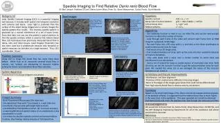
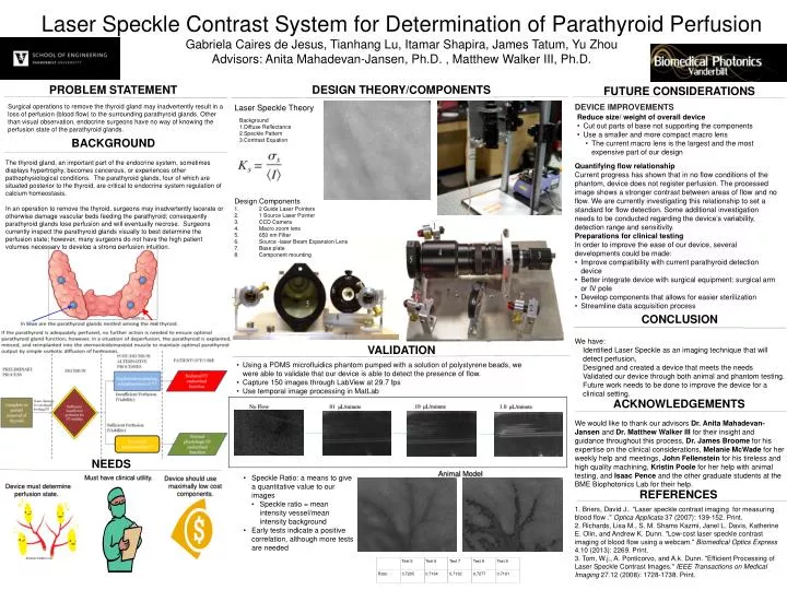
Laser Speckle Contrast System for Determination of Parathyroid Perfusion Gabriela Caires de Jesus, Tianhang Lu, Itamar Shapira , James Tatum, Yu Zhou Advisors: Anita Mahadevan -Jansen, Ph.D. , Matthew Walker III, Ph.D. .10 μ L/minute. No Flow. .01 μ L/minute. 1.0 μ L/minute.

E N D
Laser Speckle Contrast System for Determination of Parathyroid Perfusion Gabriela Caires de Jesus, Tianhang Lu, ItamarShapira, James Tatum, Yu Zhou Advisors: Anita Mahadevan-Jansen, Ph.D. , Matthew Walker III, Ph.D. .10 μL/minute No Flow .01 μL/minute 1.0 μL/minute PROBLEM STATEMENT Design Theory/Components Future Considerations DEVICE IMPROVEMENTS Surgical operations to remove the thyroid gland may inadvertently result in a loss of perfusion (blood flow) to the surrounding parathyroid glands. Other than visual observation, endocrine surgeons have no way of knowing the perfusion state of the parathyroid glands. Laser Speckle Theory • Reduce size/ weight of overall device • Cut out parts of base not supporting the components • Use a smaller and more compact macro lens • The current macro lens is the largest and the most expensive part of our design • Background • Diffuse Reflectance • Speckle Pattern • Contrast Equation BACKGROUND The thyroid gland, an important part of the endocrine system, sometimes displays hypertrophy, becomes cancerous, or experiences other pathophysiological conditions. The parathyroid glands, four of which are situated posterior to the thyroid, are critical to endocrine system regulation of calcium homeostasis. In an operation to remove the thyroid, surgeons may inadvertently lacerate or otherwise damage vascular beds feeding the parathyroid; consequently parathyroid glands lose perfusion and will eventually necrose. Surgeons currently inspect the parathyroid glands visually to best determine the perfusion state; however, many surgeons do not have the high patient volumes necessary to develop a strong perfusion intuition. • Quantifying flow relationship • Current progress has shown that in no flow conditions of the phantom, device does not register perfusion. The processed image shows a stronger contrast between areas of flow and no flow. We are currently investigating this relationship to set a standard for flow detection. Some additional investigation needs to be conducted regarding the device’s variability, detection range and sensitivity. • Preparations for clinical testing • In order to improve the ease of our device, several developments could be made: • Improve compatibility with current parathyroid detection device • Better integrate device with surgical equipment: surgical arm or IV pole • Develop components that allows for easier sterilization • Streamline data acquisition process • Design Components • 2 Guide Laser Pointers • 1 Source Laser Pointer • CCD Camera • Macro zoom lens • 650 nm Filter • Source -laser Beam Expansion Lens • Base plate • Component mounting Conclusion hormone.org We have: • Identified Laser Speckle as an imaging technique that will detect perfusion, • Designed and created a device that meets the needs • Validated our device through both animal and phantom testing. Future work needs to be done to improve the device for a clinical setting. • We would like to thank our advisors Dr. Anita Mahadevan-Jansen andDr. Matthew Walker III for their insight and guidance throughout this process,Dr. James Broome for his expertise on the clinical considerations, Melanie McWadefor her weekly help and meetings, John Fellensteinfor his tireless and high quality machining, Kristin Poole for her help with animal testing, and Isaac Penceandthe other graduate students at the BME Biophotonics Lab for their help. • 1. Briers, David J.. "Laser speckle contrast imaging for measuring blood flow ." OpticaApplicata 37 (2007): 139-152. Print. • 2. Richards, Lisa M., S. M. Shams Kazmi, Janel L. Davis, Katherine E. Olin, and Andrew K. Dunn. "Low-cost laser speckle contrast imaging of blood flow using a webcam." Biomedical Optics Express 4.10 (2013): 2269. Print. • 3. Tom, W.j., A. Ponticorvo, and A.k. Dunn. "Efficient Processing of Laser Speckle Contrast Images." IEEE Transactions on Medical Imaging 27.12 (2008): 1728-1738. Print. Validation • Using a PDMS microfluidics phantom pumped with a solution of polystyrene beads, we were able to validate that our device is able to detect the presence of flow. • Capture 150 images through LabView at 29.7 fps • Use temporal image processing in MatLab Acknowledgements Needs Animal Model • Speckle Ratio: a means to give a quantitative value to our images • Speckle ratio = mean intensity vessel/mean intensity background • Early tests indicate a positive correlation, although more tests are needed Must have clinical utility. Device should use maximally low cost components. References Device must determine perfusion state. Iheartguts.wordpress.com