Download
1 / 109
1.12k likes | 1.56k Views
ENT Procedures Operative Sequence. Myringotomy with Ear Tubes. Myringotomy. Define the procedure : A small incision is made in the tympanic membrane to allow the drainage of fluid from the middle ear and the placement of ear tubes. Myringotomy. Overall Purpose of Procedure :

E N D
ENT ProceduresOperative Sequence Myringotomy with Ear Tubes
Myringotomy • Define the procedure: • A small incision is made in the tympanic membrane to allow the drainage of fluid from the middle ear and the placement of ear tubes.
Myringotomy • Overall Purpose of Procedure: • Relieve effusion. • Effusion = ‘s fluid in the middle ear causing painful ear infections (a.k.a. acute otitis media) • Acute otitis media - Inflammation of the middle ear in which there is fluid in the middle ear accompanied by signs or symptoms of ear infection: a bulging eardrum usually accompanied by pain; or a perforated eardrum, often with drainage of purulent material (pus).
Ear Tubes • Ear tubes are tiny cylinders placed through the ear drum (tympanic membrane) to allow air into the middle ear. They also may be called tympanostomy tubes, myringotomy tubes, ventilation tubes, or PE (pressure equalization) tubes. • These tubes can be made out of plastic, metal, or Teflon and may have a coating intended to reduce the possibility of infection. • There are two basic types of ear tubes: short-term and long-term.
Ear Tubes cont. Short-term tubes are smaller and typically stay in place for six months to a year before falling out on their own. Long-term tubes are larger and have flanges that secure them in place for a longer period of time. Long term tubes may fall out on their own, but removal by an ENT surgeon is often necessary.
Myringotomy • Wound Classification: 2
Operative Sequence • 1- Incision • 2- Hemostasis • 3- Dissection • 4- Exposure • 5- Procedure (Specimen Collection possible) • 6- Hemostasis • 7- Irrigation • 8- Closure • 9- Dressing Application
Myringotomy • Instrumentation: Myringotomy tray, microscope, speculum holder, micro-instruments. • Positioning: The patient is in supine position arms tucked. MD will sit for procedure. • Prepping: Surgeon preference. Hibiclense or a Betadine Prep Kit. Betadine can be placed on cotton ball to swab the ear if MD prefers. Some use no prep at all...always ask • Draping: 4 towels and a head drape. Ask about towel clips. Ask about clear incise drape.
MyringotomyBegin your Operative Sequence • Incision: made into the tympanic membrane with myringotomy blade (this step is out of order in this surgery due to the fact that we already have an opening to work through – the ear canal)
Myringotomycont. Operative Sequence • Hemostasis: none • Dissection and Exposure: ear spec and microscope • Exploration and Isolation: MD will remove ear wax (cerumen) with ear curettes and visualize the TM.
Myringotomycont. Operative Sequence • Surgical Repair/Removal/Specimen Collection: • Will make a 2mm to 3mm incision into the TM. • Fluid is suctioned out with micro-Frasier suction, #3 or #5.
Myringotomycont. Operative Sequence • Surgical Repair/Removal/Specimen Collection: • PE tube is grasped either by the MD or scrub with alligator forceps. • Placed into the incision. • Rosen needle (actually a pick) used to manipulate PE tube into correct position. • Video • http://www.youtube.com/watch?v=j_Z-ylTtRts
Myringotomycont. Operative Sequence • Hemostasis and Irrigation: • none • Closure: • Places antibiotic drops into ear canal. • Places cotton ball in ear canal.
Myringotomy • Major Arteries: • Posterior auricular artery • Labyrinthine artery • Major Veins: • Posterior auricular vein
Myringotomy • Major Nerves: • Vestibular nerve: The vestibular nerve is one of the two branches of the Vestibulocochlear nerve (the cochlear nerve being the other). It goes to the semicircular canals via the vestibular ganglion. It receives positional information.
ENT ProceduresOperative Sequence Tonsillectomy & Adenoidectomy
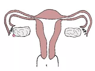
Tonsillectomy and Adenoidectomy • Tonsils are small, round pieces of tissue that are located in the back of the mouth on the side of the throat. Tonsils are thought to help fight infections by producing antibodies.
Adenoid - Lymph-like areas of tissue, or glands, that are similar to the tonsils, but they are located very high in the throat, behind the nose. They trap and filter out germs that enter the body. The adenoids also help your body fight off infection by making antibodies.
Types of Tonsils • Palatine tonsils--located on each side of the throat. • Pharyngeal tonsils--also known as adenoids are near the posterior openings of the nasal cavity. • Lingual tonsils--near the base of the tongue
You have your adenoids when you are born and they continue to grow until you are 5 to 7 years old. By school age, the adenoids begin to shrink in size, and, by the time children reach their pre-teen or teenage years, the adenoids are usually small enough to not cause any symptoms.
What Are the Symptoms of Enlarged Adenoids • A child may complain of: • difficulty breathing through the nose • is breathing through the mouth • talks as if his or her nostrils are pinched • breathes noisily • snores while sleeping • stops breathing for a few seconds while sleeping (called sleep apnea)
Tonsillectomy and Adenoidectomy • Define the procedure: removal of tissue to eradicate infection, improve the airway or remove cancer.
Tonsillectomy and Adenoidectomy • Overall Purpose of Procedure: • 3 pathological indications for removal of the tonsils and adnoids: • Infection • Hypertrophy- enlargement via cellular growth • Cancer • Pt. may suffer from tonsillitis, peritonsillar abscess, strep throat, irr. sleep patterns, difficulty swallowing. • Adenoids can become hypertrophic to the point of blocking the Eustachian tube, causing otitis media.
Tonsillectomy and Adenoidectomy • Wound Classification: 2
Operative Sequence • 1- Incision • 2- Hemostasis • 3- Dissection • 4- Exposure • 5- Procedure (Specimen Collection possible) • 6- Hemostasis • 7- Irrigation • 8- Closure • 9- Dressing Application
Tonsillectomy and Adenoidectomy • Instrumentation: T&A tray • Positioning: The patient is in supine position arms tucked. MD will sit for procedure. Spin bed 90 degrees for some Md’s. • Prepping: NONE! • Draping: 4 towels and a head drape (depends on MD). Ask about towel clips. Down sheet.
Tonsillectomy and AdenoidectomyBegin your Operative Sequence • Incision: This step comes later in the procedure. • Hemostasis: none at this point in the procedure.
Tonsillectomy and Adenoidectomycont. Operative Sequence • Dissection and Exposure: • Mouth Gag of Md choice is placed into pt’s mouth. Mouth gag WILL REST on mayo stand.
Tonsillectomy and Adenoidectomycont. Operative Sequence • Exploration and Isolation: • MD might have headlight for visualization purposes. • Tonsil is grasped with Allis clamp, possible Allis Adair. • Have FRED on mayo for dental mirror.
Tonsillectomy and Adenoidectomycont. Operative Sequence • Surgical Repair/Removal/Specimen Collection: • An incision is made in the grasped tonsil. • Incision can be made with a Snare, Laser, Curette, or Coblation Wand. • Tonsil is removed with the device of choice. • Scrub needs to have a chromic suture ready on the table for heavy bleeding.
Tonsillectomy and Adenoidectomycont. Operative Sequence • Surgical Repair/Removal/Specimen Collection: • Coblation wand uses radiofrequency waves, instead of cautery (heat) techniques, to remove tonsils and adenoids. • Coblation Tonsillectomy: http://www.youtube.com/watch?v=KizSZuqkyBc
Tonsillectomy and Adenoidectomycont. Operative Sequence • Surgical Repair/Removal/Specimen Collection: • You will repeat the same process for the other tonsil. • Adenoid – MD will retract the palate with a red rubber catheter inserted transnasally. • Clamp end of red rubber with Kelly. • MD will use dental mirror to view adenoid tissue. • Removal with same procedure as Tonsils. • You will have specimens – ask then pass off. • May need sterile safety pin to mark specimen. (usually the safety pin will go into the right tonsil. Be sure not to put the pin into your hand – Tonsils are very dirty)
Tonsillectomy and Adenoidectomycont. Operative Sequence • Hemostasis and Irrigation: • Heavy bleeding is a possibility. Always have NACL ready on back table or mayo. • Closure: packed with strung gauze. May be soaked in viscous Lidocaine. • T and A vid • Child and Adult T&A EESEDU • NEVER BREAK YOUR TABLE DOWN
Tonsillectomy and Adenoidectomy • Major Arteries: • tonsillar and ascending palatine branches of the facial artery • Major Veins: • tonsillar veins
Tonsillectomy and Adenoidectomy • Major Nerves: • tonsillar branches of glossopharyngeal nerve
SUR 122 Tracheotomy/Tracheostomy
Tracheostomy is indicated for a patient who requires emergent or elective airway management for: • prolonged ventilator dependence • acute upper airway obstruction • chronic upper airway obstruction
Pathology for Tracheotomy or Tracheostomy • Vocal cord paralysis • Neck surgery • Trauma • Prolonged intubation • Secretion management • Cannot intubate • Stridor due to tracheal blockage • Sleep apnea
Anatomy of the Neck (From Potter PA and Perry AG: Fundamentals of nursing, ed 5, St Louis, 2001, Mosby.)
Anatomy of the Larynx Anterior view of the pharynx Posterior view of the pharynx (From Thibodeau GA and Patton KT: Anthony's textbook of anatomy and physiology, ed 17, St Louis, 2003, Mosby.)
Tracheotomy/Tracheostomy • Tracheotomy temporary opening into the trachea to facilitate breathing • Tracheostomy permanent opening of the trachea and creation of a tracheal stoma • Must place tracheal tube with either • Patient will be hooked up to a ventilator • Long term tracheostomy may eventually be able to wean off ventilator, but maintain stoma that will function as their nose did prior to surgery
Anesthesia • General • Local
Medications • Local anesthetic: Lidocaine or bupivicaine with or without epinephrine • Antibiotic irrigation
Positioning • Supine • Shoulder roll • Donut headrest • Pillow under knees • Safety strap
Prep • End of chin to midchest and bedsheet to bedsheet • Prep of choice: Duraprep, betadine scrub and/or paint
Draping • Towels • Small fenestrated sheet (Pediatric lap sheet)
Minor basin Basic pack Pediatric lap sheet Other small fenestrated sheet Blades Suture or ties of surgeon’s choice (prn) Tracheotomy tray Tracheotomy tube (Shiley) Twill tape Supplies, Equipment, Instruments