Download
1 / 39
490 likes | 1.36k Views
EXCRETION, HOMEOSTASIS & OSMOREGULATION. DEFINITIONS. Metabolism – all chemical changes occurring in a cell or living organism. Includes rxns that build up or breaks down substances. Excretion – removal of unwanted products from the body such as:

E N D
DEFINITIONS • Metabolism – all chemical changes occurring in a cell or living organism. Includes rxns that build up or breaks down substances. • Excretion – removal of unwanted products from the body such as: • Waste products of chemical rxns e.g. CO2, urea, nitrogenous waste • Excess salts & water taken in with diet • Spent/used hormones and • Drugs or other foreign substances taken in the alimentary canal and absorbed by the blood
DEFINITIONS • Defaecation – removal of faeces/undigested food during egestion in humans. • Egestion – the removal of undigested food, in the form of faeces, through the anus e.g. cellulose. • NOTE: Secretion is different from excretion as it is the release of a useful substance such as an enzyme or hormone from cells.
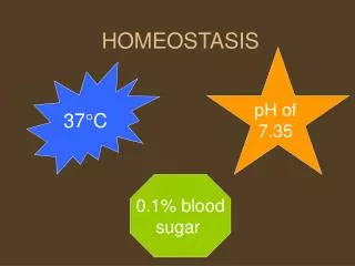
WHY IS EXCRETION IMPORTANT? • Because if these waste products remain in the organism they will become toxic or poisonous to the organism by damaging its tissues. Also, excretion is necessary to maintain a balance in the body (homeostasis).
EXCRETORY ORGANS IN ANIMALS • Name them • Lungs: remove CO2 • Kidneys: remove urea, other nitrogenous waste from the blood, excess water, salts, hormones & drugs • Liver: yellow-green pigment from the breakdown of Hb is excreted with bile into small intestine & excreted with faeces this pigment gives faeces its brown colour. Also produces urea (note pg 78 Ali) • Skin: salt & urea are excreted in sweat as a response to an increase in temperature
EXCRETORY ORGANS • What are the excretory organs in plants? • Leaves - stomata • Plants don’t need such elaborate organs like animals. Why? • Because their metabolism isn’t fast. • Why? • Immobile
EXCRETORY PRODUCTS IN PLANTS • What are some examples? • CO2 from respiration. CO2 not used by chloroplasts during photosynthesis is excreted through the stomata via diffusion. • O2 from photosynthesis. Some is recycled for respiration but unused O2 is excreted through the stomata.
Water from photosynthesis • Calcium oxalate which is stored in parts of plants such as their leaves, bark, flowers, fruits & seeds. When these parts are shed these products are removed. • Alkaloids (poisonous nitrogen compounds) are contained in milky substances of plants • Tannins are poisonous waste products stored in the bark of trees. Tannins are removed when the tree sheds its bark. Mangroves possess tannins.
PARTS OF THE EXCRETORY SYSTEM • Kidney – main organ • Size – each is 12 cm long by 7 cm wide • The left kidney is slightly higher than the right • Bean-shaped and red located at the back of the abdominal cavity (see diagram pg 129 Ali) behind the intestines. • Put hand on hips – thumbs indicate location of kidneys
Kidneys receive “dirty” blood from renal artery while the renal vein takes cleansed/filtered blood away from kidneys to the heart. • Nitrogenous & other waste flow down through the ureter to the bladder. • The ureter connects each kidney to the bladder (muscular bag @ bottom of kidney)
The bladder is drained by the urethra to the opening of penis/vagina. • At the top of the urethra are 2 sets of sphincter muscle which controls the release of urine. The lower sph. muscle is controlled voluntarily while the upper muscle is controlled involuntarily. It automatically relaxes when the bladder is full. • When the bladder is full the stretching stimulates sensory nerve endings in its wall which sends nerve impulses to the brain causing urine to be released. This is called urination. • Babies can’t control their voluntary sph. muscles
KIDNEY STRUCTURE Three main parts: • Cortex • Medulla - pyramids • (Nephrons) • Pelvis
KIDNEY STRUCTURE • Contains about 160km of blood vessels & more than a million nephrons/kidney tubules • Two areas: darker outer region – cortex; paler inner region – medulla • Cortex contains many tiny blood vessels which branch from renal artery. • Cortex also contains nephrons which run through the medulla.
Medulla is connected to ureter (Fig. 12.3 Ali) • Medulla consists of several cone-shaped areas called pyramids which point inwards towards the concave side of the kidney i.e. into the pelvis • Urine drains from tips of pyramids (at the pelvis) into funnel-shaped spaces formed by the top of ureter • Ureter carries urine to the bladder
http://www.tutorvista.com/content/biology/biology-ii/excretion-and-osmoregulation/excretory-system.phphttp://www.tutorvista.com/content/biology/biology-ii/excretion-and-osmoregulation/excretory-system.php • http://www.medindia.net/animation/anatomy_urinary.asp
STRUCTURE OF A NEPHRON • Basic unit of the kidney • Bowman’s capsule - This closed end at the beginning of the nephron is located in the cortex. • Glomerulus – made up of Bowman’s capsule and glomerular capillaries • Proximal convoluted tubule - The first twisted region after the Bowman's capsule; it's in the cortex. • Loop of Henle - A long, hairpin loop after the proximal tubule, it extends from the cortex down into the medulla and back.
Distal/Second convoluted tubule - This second twisted portion of the nephron after the loop of Henle is located in the cortex. • Collecting duct - This long straight portion after the distal tubule that is the open end of the nephron extends from the cortex down through the medulla.
STRUCTURE OF A NEPHRON • Kidney has millions of nephrons/kidney tubules – 3cm long • They begin in the cortex, loops down into the medulla, back to cortex, down through the medulla to the pelvis where they join with the ureter • The 1st part of the nephron is the Bowman’s capsule which is a round, cup-shaped object which is ~ 0.2mm in diameter. • Each capsule encloses a ball of finely divided and inter-twined blood capillaries called the glomerulus (plural glomeruli).
The glomeruli are supplied with blood containing N2 and other wastes products to be cleansed. • Each tubule/nephron emerges from the Bowman’s capsule on the side opposite to the glomerulus, goes into a series of loops, then joins the wider collecting duct. • These ducts collect urine from the nephrons and transport it straight trough the medulla to the tips of the kidneys
NOTE: Blood supply to nephrons • Oxygenated blood comes to the kidneys at a high pressure through the renal artery from the main aorta. • Inside the kidney, the renal artery divides into arterioles (afferent arteriole) which carry blood to glomeruli capillaries with a small drop in pressure • The blood vessel (efferent arteriole) which leaves each glomerulus branches into a capillary network around the coiled and looped part of each nephron, before joining the renal vein.
The afferent arteriole has a larger diameter than the efferent arteriole to withstand the pressure of blood entering it. • This difference in arteriole diameters helps to raise the blood pressure in the glomerulus.
FORMATION OF URINE • Urine is formed in 2 stages: • Blood is filtered into the kidney tubules to form a clear liquid (filtrate) which contains the waste urea and many useful substances which the body can’t lose. This stage is called filtration. • These useful substances are reabsorbed from the filtrate back into the blood leaving urea and other useless substances in the kidney tubules. This stage is called reabsorption.
Pressure/Ultra-filtration • Filtration of the blood occurs in the Bowman’s capsule through 2 layers of membranes: the capillary wall of each glomerulus & the inner wall of each Bowman’s capsule. • These membranes have a surface area of 1 m2
Blood enters the Bowman’s capsule at a high pressure due to the difference in diameters of the afferent & efferent arterioles. • This pressure allows small molecules such as water, glucose, AA, vitamins, hormones, salts and urea to pass into the Bowman’s capsule. • Together they form the liquid glomerular filtrate. • Larger molecules – plasma proteins & blood cells remain in the blood outside the B. capsule.
The filtering process in kidneys is similar to the formation of tissue fluid as both processes involve blood being forced at high pressure through capillary walls where proteins and blood cells (erythrocytes & leucocytes) are filtered out leaving a clear liquid. • The glomerular filtrate formed in the B. capsule drains into another part of the nephron: the proximal convoluted tubule. • Kidneys produce 125cm3 of g. filtrate a minute
2. Selective reabsorption • This occurs via three mechanisms: • Osmosis • Diffusion, and • Active Transport. • Occurs in the proximal convoluted tubule. • Most of the volume of the filtrate solution is reabsorbed in the proximal convoluted tubule (PCT) • This prevents dehydration as useful substances are reabsorbed into the capillaries that surround each nephron.
Glucose, water and some salts need to be kept in the blood. • Why is glucose reabsorbed? • Glucose is used for energy. • Most of the energy consumed by the kidneys is used in the reabsorption of sodium ions (Na+), which are solutes - that is, they are dissolved in the water component of the filtrate solution. As the concentration of Na+ in the filtrate solution is high (about the same as the concentration of Na+ in blood plasma), Na+ moves from the tubular fluid into the cells of the PCT
All glucose, amino acids and 85% of mineral ions are reabsorbed by active transport. Small proteins are reabsorbed. 80% of water is absorbed back into the blood by osmosis. The proximal convoluted tubule cells have many mitochondria to provide ATP for active transport and microvilli to increase surface area for absorption. • Other such substances that are reabsorbed with Na+ via active transport include glucose, amino acids, lactic acid, and bicarbonate ions (HCO3-). These then move on through cells via diffusion and/or other transport processes
OSMOREGULATION • This is the regulation of body fluids. • Another role of kidneys • Kidneys can control the concentration of urine & thus regulate the water content of the blood. • What will the kidneys do if one were to drink a litre of water all at once? • The kidneys will respond to this imbalance by producing a larger volume of dilute urine.
What will happen if the blood is concentrated? • The kidneys will produce a smaller volume of urine. • How can the blood become concentrated? • Diet – salty, a lot of sugar. • These changes are controlled by a hormone produced by the pituitary gland at the base of the brain. • Hormone: anti-diuretic hormone/ADH or vasopressin.
ADH begins to work when your body loses too much water e.g. sweating or not replacing water loss via drinking. • Loss of water = increase of concentration blood. • This is detected by special cells in the hypothalamus which is a region of the brain. • These cells are sensitive to the solute concentration of blood which causes the pituitary gland to release more ADH. • ADH travels through the blood stream to the kidney. • ADH causes the walls of the collecting ducts to become more permeable to water to allow more water to be absorbed back into the blood..
This makes the urine concentrated so the body loses less water but makes the blood dilute. • When the water content of blood returns to normal – ADH is no longer released. • Kidney tubules reabsorb less water. • If a person drinks too much water, blood is too dilute – less ADH is secreted • Leads to the kidney tubules becoming less permeable to water – more water passes out urine. • Hence ADH ensures that the internal environment is kept constant.
ADH & Negative feedback • Internal environment • Change in internal environment • Detector senses change • Correcting action • Internal environment returns to normal Negative feedback loop switches off cause of correcting action
To review as well • Kidney failure • Homeostasis • Osmoregulation in fish, land animals e.g. kangaroo rat which has a VERY long loop of Henle, amoeba, plants • Liver & its functions w.r.t. homeostasis • Controls amount of sugar in blood • Bile production • deamination • Gallstones • Skin- excretory & homeostatic organ
Nephron activity: http://www.sumanasinc.com/webcontent/animations/content/kidney.html • http://www.youtube.com/watch?v=glu0dzK4dbU&feature=related
REFERENCES • file:///G:/Form%204/Biology/Kidneys%20etc/kidney%5B1%5D.ppt#256,1,Excretion and the Kidneys • http://www.biologie.uni-hamburg.de/b-online/library/falk/Excretion/excretion.htm • http://www.sumanasinc.com/webcontent/animations/biology.html • http://kidney.niddk.nih.gov/Kudiseases/pubs/yourkidneys/ Kidney stone video • http://health.howstuffworks.com/kidney.htm • **http://videos.howstuffworks.com/hsw/25407-human-body-the-urinary-system-video.htm • Kids - http://kidshealth.org/kid/htbw/kidneys.html# • Teens - http://kidshealth.org/teen/your_body/body_basics/kidneys.html# • Useful Nephron video - http://www.youtube.com/watch?v=glu0dzK4dbU&feature=related • Urine formation complicated - http://www.youtube.com/watch?v=AdlfxBooqIA&feature=related • Good one (~20mins) - http://videos.howstuffworks.com/hsw/20550-human-body-systems-the-excretory-system-video.htm • Diagrams - http://academic.kellogg.cc.mi.us/herbrandsonc/bio201_McKinley/Urinary%20System.htm • Interesting - http://www.uic.edu/classes/bios/bios100/lecturesf04am/lect21.htm • http://www.daviddarling.info/encyclopedia/K/kidney.html accessed May 27, 2009 • http://www.ivy-rose.co.uk/Topics/Urinary_System_Kidneys_Actions.htm accessed June 3, 2009 Ultra filtration & absorption info • Gallstones http://digestive.niddk.nih.gov/ddiseases/pubs/Gallstones/ Accessed June 8, 2009 • Gallstones http://www.emedicinehealth.com/gallstones/article_em.htm Accessed June 8, 2009 • Gallstone image: http://www.hipusa.com/webmd/encyclopedia/gallstones/index.html • http://kidshealth.org/teen/question/illness_infection/hypoglycemia.html# Accessed June 8, 2009 • http://www.medicinenet.com/hypoglycemia/article.htm Accessed June 8, 2009 • Kidney http://www.iol.co.za/matric_matters_pdfs/vol24pdf/vol-24_page12.pdf Accessed June 8, 2009 • http://www.aqavic.org.au/sci_facts/neurogenic_bladder.html Accessed 9 March 2011 • http://www.nutritionreview.org/library/bladder_control.php