Download
1 / 13
160 likes | 549 Views
Gametogenesis. the formation of gametes in animals although Gametogenesis in males and females is generally the same there are some general differences. p. 170, Fig 5.18. Spermatogenesis. Oogenesis. 1˚ Spermatocyte (2n). 1˚ Oocyte (2n). Meiosis I. 2˚ Spermatocytes (n). 2˚ Oocyte

E N D
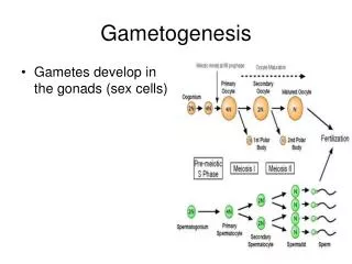
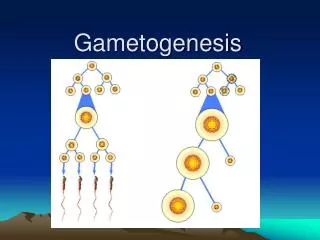
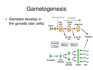
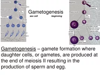
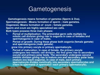
Gametogenesis the formation of gametes in animals although Gametogenesis in males and females is generally the same there are some general differences
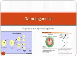
p. 170, Fig 5.18 Spermatogenesis Oogenesis 1˚ Spermatocyte (2n) 1˚ Oocyte (2n) Meiosis I 2˚ Spermatocytes (n) 2˚ Oocyte (n) Meiosis II Spermatids (n) Ootid (n) Polar Bodies (n) Spermatozoa (n) Ovum (n)
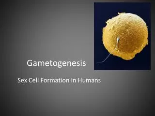
Males equal division of cytoplasm since sperm cells are streamlined for movement, they cannot carry excess weight make more sex cells than females Females cytoplasm does not divide equally after each division one daughter cell called the ootid receives most of the cytoplasm uses the extra cytoplasm and organelles for future cell divisions (if fertilization) the other cells called polar bodies die only one ovum (egg cell) is produced by meiosis Males vs. Females“Battling Sexes”
Males the diploid spermatocytes - the cells which give rise to sperm cells - are capable of many mitotic cell divisions before meiosis ever begins can produce one billion sperm cells everyday Females baby females have two million primary oocytes in their ovaries most are absorbed into the body of this number 400 to 500 will be released during the reproductive years primary oocytes have already entered meiosis I remained suspended in prophase I until the female reaches reproductive age or puberty Males vs. Females“by the Numbers”
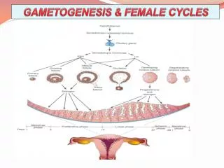
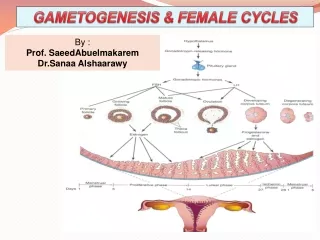
Menstrual Cycling starting at the first menstrual cycle, meiosis will resume in one oocyte at a time, once a month until menopause menopause is the end of menstrual cycling in women usually occurs between the ages of 40 to 55 the remaining oocytes left in the ovaries are nonfunctional or unresponsive and are no longer released
Chromosomes All chromosomes in females are found as homologous pairs Not all chromosomes in males are found as homologous pairs
There is one different pair of chromosomes in males and females. In females the pair is always two rod shaped chromosomes (XX) In males there is one rod-shaped chromosome and one hook-shaped chromosome (XY) These chromosomes are called Sex Chromosomes Chromosomes that are not sex chromosomes are calledAutosomes
Abnormal Meiosis Like most processes sometimes meiosis makes mistakes eg. Nondisjunction • occurs when two homologous chromosomes move to the same pole during meiosis • the result is one daughter will be missing a chromosome (22) and the other will have an extra chromosome (24) • cells that lack or have too much genetic information will not function properly Aside: Nondisjunction can also happen during mitosis, but the effects are less devastating than during meiosis. WHY?
Abnormal Meiosis Trisomy • the condition where there arethree homologous chromosomes in place of a homologous pair • zygote with 47 chromosomes • eg. Down Syndrome or Klinefelter Syndrome Monosomy • the condition where there is a single chromosome in place of a homologous pair • zygote with 45 chromosomes • eg. Turner Syndrome
Karyotyping Karyotype Chart a picture of chromosomes arranged in homologous pairs in descending order by size, with sex chromosomes placed last How? 1. mix a small amount of tissue with a solution that stimulates mitotic division 2. add another solution which stops the division at metaphase (the best phase to karyotype since the chromosomes are the most condensed) 3. the metaphase cells are placed on a slide and stained so that distinctive bands appear 4. a photograph is taken and blown up and then the individual chromosomes are cut out and matched with its homologous pair.