Download
1 / 11
150 likes | 476 Views
Areas of Auscultation Chapter 5. Ara G. Tilkian, MD, FACC Instructor Patricia L. Thomas, MBA, RCIS. Outline. Traditional Areas of Auscultation Left Ventricular Area Right Ventricular Area Left Atrial Area Right Atrial Area Aorta Area Pulmonic Area. Traditional Areas of Auscultation.

E N D
Areas of AuscultationChapter 5 Ara G. Tilkian, MD, FACC Instructor Patricia L. Thomas, MBA, RCIS
Outline • Traditional Areas of Auscultation • Left Ventricular Area • Right Ventricular Area • Left Atrial Area • Right Atrial Area • Aorta Area • Pulmonic Area
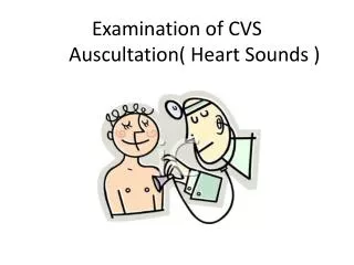
Traditional Areas of Auscultation • Mitral Area –Cardiac Apex • Tricuspid Area-4th & 5th intercostal space of left sternal border • Aortic Area-2nd intercostal space of the right sternal border • Pulmonic Area-2nd intercostal space left sternal border
Left Ventricular Area • Aortic component of the 2nd heart sound A2 • Cardiac Apex and extends laterally in the 3rd and 4th interspaces to left sternal border in one direction & to the anterior axillary line in the other.
Right Ventricular Area • Tricuspid Stenosis or regurgitation, pulmonary regurgitation and ventricular septal defect.
Left Atrial Area • Mitral Regurgitation • Anterior chest-listen left of apex • Posterior left thorax-area between the spine and the border of the scapula at the level of its tip
Right Atrial Area • Tricuspid insufficiency is best heard at the right sternal border in the 4th and 5th interspaces.
Aortic Area • Aortic Ejection Click and A2 • Aortic stenosis & Insufficiency • Increased aortic flow • Dilatation on the ascending aorta • Abnormalities of the carotid or subclavian arteries
Pulmonic Area • Pulmonic Ejection & P2 • Pulmonary Stenosis & insufficiency • Increased pulmonary flow & stenosis • Patent Ductus arteriosus PDA
THE ENDOFCHAPTER 5 Tilkian, Ara MD Understanding Heart Sounds and Murmurs, Fourth Edition, W.B. Sunders Company. 2002, pp. 43-48.