Download

1 / 52
550 likes | 796 Views
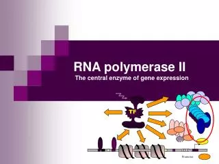
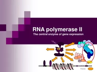
TF. TBP. TATA. Promoter. RNA polymerase II The central enzyme of gene expression. Enzymatic function. Enzymatic reaction: NTP RNA + PPi (1969) RNA n + NTP + (Mg ++ + templat) = RNA n+1 + PP i Processive - can transcribe 10 6 bp template without dissociation

E N D
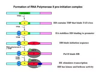
TF TBP TATA Promoter RNA polymerase II The central enzyme of gene expression
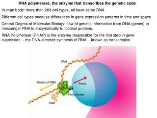
Enzymatic function • Enzymatic reaction: NTP RNA + PPi (1969) • RNAn + NTP + (Mg++ + templat) = RNAn+1 + PPi • Processive - can transcribe 106 bp template without dissociation • mRNA levels can vary with a factor of 104 • Central role : unwind the DNA doble helix, polymerize RNA, and proofread the transcript • RNAPII assembles into larger initiation and elongation complexes, capable of promoter recognition and response to regulatory signals
Polymerization reaction • 1. Initiation • PIC assembly (pre-initiation complex) • Open complex formation • Promoter clearance • 2. Elongation - transition to stable TEC • (transcription elongation complex) • 3. Termination
Subunit structure • Composition and stochiometry • 12 polypeptides • 2 large (220 and 150 kDa) + 10 small (10 - 45 kDa) • Stoichiometry: 1, 2 and <1 • Yeast: 10 essensial, 2 non-essensial • Phosphorylated subunits: RPB1 and RPB 6 • Highly conserved between eukaryotes • Several subunits in yeast RNAPII can be functionally exchanged with mammalian subunits
Subunits of RNA polymerase II • The yeast model
Evolutionary conservation of Subunits of RNA polymerase II • Core-enzyme with the active site • RPB1 (´-like) binds DNA • RPB2 (-like) binds NTP • RPB3 and RPB11 (-like) assembly factors • Evolutionary conserved mechanism of RNA synthesis • Common subunits • RPB5, 6, 8, 10 and 12 common to RNAPI, II and III • Common functions? • Ulike prokaryotic RNAP, the eukaryotic RNAPII is unable by itself to recognize promoter sequences Prokaryotic ´ DNA-binding NTP-binding Eukaryotic
Structure 1999 - 2001 Taq RNAP (open) yRNAPII (closed) Arm (ß2) open NTP Arm closed ASC Jaws Jaws • 1999: First 3D-structure published • Yeast yRNAPII 6Å resolution • yRNAPII + DNA/RNA low resolution • Taq RNAPII high resolution 3.3Å • • 2000: 3D crystal - high resolution • 10 subunit yRNAPII - 3Å resolution • • 2001: 3D crystal - higher resolution • High-resolution structures of two conformationally • distinct forms of yRNAPII; and an active • yRNAPII trx elongation complex (TEC). DNA upstream DNA nedstrøms ASC = active site channel
Yeast RNAPII • The two largest subunits, Rpb1 and Rpb2, form masses with a deep cleft between them • The small subunits are arranged around
Overall Structure • A helix of Rpb1 bridges the cleft, and the carboxy-terminal region of Rpb2 extends to the opposite side • The Rpb1-Rpb2 complex is anchored at one end by a subassembly of Rpb3, Rpb10, Rpb11 and Rpb12 • Mg2+ occurs within a loop of Rpb1 • B-form DNA lies in the Rpb1-Rpb2 cleft • About 20 bp from the edge of the polymerase to the active site
Several important subdomains • Channel for DNA template (downstream) • Jaws • Clamp • Wall • Active site • Pore for NTP entry • Channel for RNA exit • Hybrid melting • fork loop 1 + rudder + lid • Dock • CTD
Channel for DNA template: 25Å channel through the enzyme yRNAPII
Jaws • A pair of jaws that appear to grip DNA downstream of the active center. • Rpb5 and regions of Rpb1 and Rpb9 forms ”jaws” that appear to grip the DNA • Both the upper and lower jaw may be mobile, opening and closing on the DNA • The larger NH2-terminal domain of Rpb5 can move
A clamp retains DNA • A clamp on the DNA nearer the active center may be locked in the closed position by RNA great stability of complexes. • The ”clamp” = N-terminal regions of Rpb1 and Rpb6, and the C-terminal regions of Rpb2 • This binding site is important for the great stability of a transcribing complex and processivity of transcription >30Å move
A clamp retains DNA Cramer 04
Moving through the compartments • DNA enters RNAPII in the first chamber (jaw-lobe module). • This module binds 15–20 bp of the downstream DNA without melting it.
Moving through the 2. compartment • The DNA melts as it enters the second chamber • a 27-40 Å cleft that contains the active site near the point of DNA melting. • The first 8–9 nt of product RNA form a heteroduplex with the template DNA (hybrid). • At the upstream end, a wall of protein blocks extension of the RNA:DNA hybrid
A wall blocks the path DNA Cramer 04
The active site • Reaction catalyzed • Two NTP sites: A + E • Addition site • Entry stie seminar Boeger, H., Bushnell, D.A., Davis, R., Griesenbeck, J., Lorch, Y., Strattan, J.S., Westover, K.D. and Kornberg, R.D. (2005) Structural basis of eukaryotic gene transcription. FEBS Lett, 579, 899-903.
A funnel for substrate entry • A pore in the protein complex beneath the active center may allow entry of substrates for polymerization.
The wall and the DNA-RNA hybrid site • Transcribing polymerases have a DNA-RNA hybrid of 8-9 bp in an unwound region of DNA, with the growing end of RNA at the active site • The DNA-RNA hybrid can’t get longer because of an element from Rpb2 that is blocking the path • Because of this ”wall”, the DNA-RNA hybrid must be tilted relative to the axis of the downstream DNA • At the upstream end of the DNA-RNA hybrid, the strands must separate
RNA-DNA hybrid - 90o • The DNA is unwound, with 9 bp of DNA–RNA hybrid in the active center region. • The axis of the hybrid helix is at nearly 90o to that of the entering DNA duplex, due to the wall. Westover, K.D., Bushnell, D.A. and Kornberg, R.D. (2004) Structural basis of transcription: nucleotide selection by rotation in the RNA polymerase II active center. Cell, 119, 481-489.
Melting the RNA-DNA hybrid • Melting of the DNA–RNA hybrid due to the intervention of three protein loops: • Rudder (”ror”) contacting DNA, and • Lid - contacting RNA. A Phe side chain serves as a wedge to maintain separation of the strands. • Fork loop 1 contacts base pairs 6 and 7, limiting the strand separation. • The three loops form a strand-loop network, whose stability must drive the melting process. Westover, K.D., Bushnell, D.A. and Kornberg, R.D. (2004) Structural basis of transcription: nucleotide selection by rotation in the RNA polymerase II active center. Cell, 119, 481-489.
RNA exit • Groove in the RNAPII structure for RNA exit. • Length and localication of the groove are appropriate for binding a region of RNA 10-20 nt from the active site. • RNA in the groove at the base of the clamp could explain the great stability of transcribing complexes Westover, K.D., Bushnell, D.A. and Kornberg, R.D. (2004) Structural basis of transcription: nucleotide selection by rotation in the RNA polymerase II active center. Cell, 119, 481-489.
Dock • Contact region for speficic interating GTFs • More next lecture Cramer 04
Rbp7/4 - recently determined Cramer 04
Opening and closing of RNAPII • Open RNAP during formation of PIC • moderate stability • Extended footprint - DNA folded around the enzyme • Strand separation and placement of template in active site, transcription bubble • ”Abortive initiation” (RNA up to 10 nt) without structural change • RNAP closes during promoter clearance and transition to TEC • contacts to PIC are disrupted and new contacts with elongation factors formed • CTD is phosphorylated (more later) • Conformational change to a ternary complex of high stability • Closed chanel around the DNA-RNA hybrid in the active site • RNAP opens and becomes destabilised during termination • Reversal of the structural changes - opening and destabilization • Prokaryot: RNA-hairpin opens RNA exit - destabilization buble - AU-rich dissociation
Conformational changes during the transcription cycle Destabilised again druing termination open open Rearranged to a very stable TEC (transcription elongation complex) that can move trough 104-106 bp without dissociation. Footprint reduced (35 bp). Euk: phosphoryl. of CTD and association with elong.factors Large footprint (70-90 bp) caused by DNA wrapped around RNAP ”Abortive initiation” may happen in this state (RNA <10 nt) closed open Transition
CTD - C-terminal domain • Conserved tail on the largest subunit: (YSPTSPS)n • Yeast n = 26, humans n = 52 • hydrophilic exposed tail • Unique for RNAPII • Essential function in vivo • >50% lethal • partial deletions cause conditional phenotype • Truncations impairs enhancer functions, initiation, and mRNA processing. • Mice with 2x ∆13 CTD: high neonatal lethality + born smaller • Different promoters show different dependence on CTD • yeast CTD-deletion n=2711, effect: GAL4 HIS4=
P P P P P P P CTD is highly phosphorylated • Full of residues that can be phosphorylated • Tyr-Ser-Pro-Thr-Ser-Pro-Ser • Reversible phosphorylation occurs on both Ser and Tyr • Creates different forms of RNAPII • RNAPIIO - hyperphosphorylated (Mr=240k) • ≈ 50 phosphates (one per repeat) • Abl- phosphorylated in vitro ≈30 fosfat • RNAPIIA - without phosphate (Mr=214k) • RNAPIIB - with CTD deleted
CTDs phosphorylation changes during the transcription cycle • Function of RNAPIIA ≠ RNAPIIO • PIC assembly: only non-phosphorylated RNAPIIA • Elongation complex: only hyperphosphorylated RNAPIIO • Phosphorylation status changes during the transcription cycles • Phosphorylation occurs after PIC assembly • dephosphorylation - on free polymerase or upon termination
P P P P P P P P P P P P P P CTD - properties = phosphorylation + protein binding
CTD is also binding several proteins • SRBs - supressors of RNA pol. B • genetic evidence • mutated SRB proteins may abolish the effect of CTD deletions • SRBs = components of the Mediator - more later • GTFs • TBP • TFIIF (74 kDa subunit) • TFIIE (34 kDa subunit) • Several proteins involved in pre m-RNA processing • Many CTD-binding proteins have been identified having important functions in splicing and termination - more later
CTD structure? • CTD peptide structure • is shown as a coil, with alternating β-turns (cyan) and extended regions (pink). • Seminar Meinhart, A. and Cramer, P. (2004) Recognition of RNA polymerase II carboxy-terminal domain by 3'-RNA-processing factors. Nature, 430, 223-226.
CTDs function • 1. Function: in initiation - recruitment • Role in recruitment of RNAPII to promoters • Only RNAPIIA can initiate PIC-assembly • Interactions with GTFs (more next lecture) • 2. Function: in promoter clearance • Def: The process whereby RNAPII undergoes the transition to hyperphosphorylated elongation modus • Hypothesis: CTD phosphorylation disrupts interactions and RNAPII gets free from PIC • Hypothesis: CTD phosphorylation creates novel interactions with elongation factors
P P P P CTDs function cont • 3. Function: during elongation • Regulation during elongation • Trx of several proto-oncogenes such as myc, myb, fos regulated via pausing (block of elongation) • Tight coupling : transcription - pre-mRNA processing • More later Pause
Regulation by CTD kinases/ phosphatases - the logic • CTD kinases • specific for free RNAPII repression • specific for assembled RNAPII activation • CTD phosphatases • specific for free RNAPII activation • specific for template associated RNAPII repression
CTD kinases • Several CTD-kinases = Cdk´s • Four of the putative CTD kinases are members of the cyclin-dependent kinase (CDK)/cyclin family whose members consist of a catalytic subunit bound to a regulatory cyclin subunit.
Candidate CTD-kinases • CTD kinase in TFIIH - positive action (more in next lecture) • Good candidate with respect to timing and location • A multisubunit factor recruited in the last step of PIC assembly • TFIIH associated CTD-Kinse = MO15/CDK7 (vertebrates) = KIN28 (yeast) • Phosphorylates Ser5 in CTD • CTD kinase Srb10/11 - negative action • cyclin-cdk pair (SRB10/11) • Conserved - human SRB10/11 also called CDK8-cyclin C • Isolated as a ∆CTD supressor - but recessive and with negative function in trx • Phosphorylates Ser5 in CTD • Unique by phosphorylating CTD of free RNAPII - hence negative effect on trx • Other candidates • in vitro - CTD is substrate for several kinases • CDK9: component of P-TEFb, a positive-acting elongation factor • MAP kinases (ERK type), • c-Abl Tyr-kinase, CDK7 CDK8 CDK9 - Srb10/11 RNAP IIO (Y1S2P3T4S5P6S7)n + TFIIH (Kin28)
RNAP IIO RNAP IIO (Y1S2P3T4S5P6S7)n (Y1S2P3T4S5P6S7)n Pattern of serines phosphorylated changes during the transcription cycle • Recent evidence suggests that the phosphorylation pattern changes during transcription • Ser 5 phosphorylation is detected mainly at promoter regions (initiation) • Ser 2 phosphorylation is seen only in coding regions (elongation) Initiation Elongation P P
RNAP IIO RNAP IIO (Y1S2P3T4S5P6S7)n (Y1S2P3T4S5P6S7)n Pattern of serines phosphorylated changes during the transcription cycle • Recent evidence suggests that the phosphorylation pattern changes during transcription • Ser 5 phosphorylation is detected mainly at promoter regions (initiation) • Ser 2 phosphorylation is seen only in coding regions (elongation) Initiation Elongation P P
May different promoters recruit different CTDK? • Evidence for distinct phosphorylatedforms • Both Ser and Tyr phosphorylated - different kinases • Several activities in vitro • CTK1 disruption in yeast reduces but don’t abolish CTD phosphorylation • If different CTDKs are recruited at different promoters promoter-imprinting ! • Multiple CTD kinases may be caused by • redundancy • different promoters recruit different kinases • different timing / cell cycle • different subcellular localization
Complex network- Links to cell cycle CDK8 CDK7 CDK9
CTD phosphatases • First CTD phosphatase characterized = FCP1 • Fcp1p is necessary for CTD dephosphorylation in vivo • yeast cells with temperature-sensitive mutations have severe defects in poly(A)+ mRNA synthesis at the nonpermissive temperature • FCP1 dephosphorylates Ser2 in CTD • Function - elongation and recycling • human FCP1 can stimulate elongation by RNAPII • FCP1 presumably helps to recycle RNAP II at the end of the transcription cycle by converting RNAP IIO into IIA for another round of transcription. • Other CTD phosphatases specific for Ser5 • SCPs - a family of small CTD phosphatases that preferentially catalyze the dephosphorylation of Ser5 within CTD. Expression of SCP1 inhibits activated transcription from a number of promoters. SCP1 may play a role in transition from initiation/capping to processive transcript elongation. • Ssu72, a component of the yeast cleavage/polyadenylation factor (CPF) complex, is a CTD phosphatase with specificity for Ser5-P. Ssu72 may have a dual role in transcription: in recycling of RNAP II and in trx termination.
CTD phosphatase • FCP1 is phosphoryolated - regulatory target? • FCP1 is phosphorylated at multiple sites in vivo. Phosphorylated FCP1 is more active in stimulating transcription elongation than the dephosphorylated form. • CTD phosphatase probably under regulation • Ex: The peptidyl-prolyl isomerase Pin1 influences the phosphorylation status of the CTD by inhibiting the CTD phosphatase FCP1 and stimulating CTD phosphorylation by cdc2/cyclin B. • Seminar: Yeo et al. (2005) Small CTD phosphatases function in silencing neuronal gene expression. Science, 307, 596-600. • FCP1 is disease related • Varon et al. (2003) Partial deficiency of the C-terminal-domain phosphatase of RNA polymerase II is associated with congenital cataracts facial dysmorphism neuropathy syndrome. Nat Genet, 35, 185-189.
Fcp1 recruited through Rbp4/7 • 3D Fcp1 Kamenski et al. (2004) Mol Cell, 15, 399-407.