Download

1 / 113
1.18k likes | 1.54k Views
NEOPLASIA. All tumors, benign and malignant, have two basic components: (1) proliferating neoplastic cells that constitute their parenchyma proliferating "cutting edge" determine their nature,

E N D
All tumors, benign and malignant, have two basic components: (1) proliferating neoplastic cells that constitute their parenchyma proliferating "cutting edge" determine their nature, (2) supportive stroma made up of connective tissue and blood vessels. growth and evolution of neoplasms are critically dependent on their stroma. adequate stromal blood supply is requisite, and the stromal connective tissue provides the framework for the parenchyma. stromal support is scant - neoplasm is soft and fleshy. parenchymal cells stimulate the formation of an abundant collagenous stroma--referred to as desmoplasia.
The nomenclature of tumors based on the parenchymal component. Benign Tumors. In general, benign tumors are designated by attaching the suffix -oma to the cell of origin. However nomenclature of benign epithelial tumors is more complex. They are variously classified, some based on their cells of origin, others on microscopic architecture, and still others on their macroscopic patterns. Adenoma benign epithelial neoplasm that forms glandular patterns as well as to the tumors derived from glands but not necessarily reproducing glandular patterns. Benign epithelial neoplasms producing microscopically or macroscopically visible finger-like or warty projections from epithelial surfaces are referred to as papillomas Those that form large cystic masses - cystadenomas. Some tumors produce papillary patterns that protrude into cystic spaces and are called papillary cystadenomas. When a neoplasm, benign or malignant, produces a macroscopically visible projection above a mucosal surface and projects - a polyp. Malignant Tumors. The nomenclature essentially follows the same schema used for benign neoplasms, with certain additions.
Malignant tumors arising in mesenchymal tissue - sarcomas Malignant neoplasms of epithelial cell origin, derived from any of the three germ layers - carcinomas. a glandular growth pattern microscopically is termed an adenocarcinoma. the organ of origin Not infrequently, a cancer is composed of undifferentiated cells and must be designated merely as a poorly differentiated or undifferentiated malignant tumor. In most neoplasms, benign and malignant, the parenchymal cells bear a close resemblance to each other divergent differentiation of a single line of parenchymal cells creates what are called mixed tumors. . The great majority of neoplasms, even mixed tumors, are composed of cells representative of a single germ layer. The teratoma, in contrast, is made up of a variety of parenchymal cell types representative of more than one germ layer, usually all three. They arise from totipotential cells and so are principally encountered in the gonads, although, rarely, they occur in sequestered primitive cell rests elsewhere. An ectopic rest of normal tissue is sometimes called a choristoma- Analogously, aberrant differentiation may produce a mass of disorganized but mature specialized cells or tissue indigenous to the particular site, referred to as a hamartoma.
CHARACTERISTICS OF BENIGN AND MALIGNANT NEOPLASMS in general, there are criteria by which benign and malignant tumors can be differentiated, and they behave accordingly: (1) differentiation and anaplasia, (2) rate of growth, (3) local invasion, (4) metastasis. Differentiation and Anaplasia These terms apply to the parenchymal cells of neoplasms. Differentiation - extent to which parenchymal cells resemble comparable normal cells, both morphologically and functionally. Well-differentiated tumors are thus composed of cells resembling the mature normal cells of the tissue of origin of the neoplasm Poorly differentiated or undifferentiated tumors have primitive-appearing, unspecialized cells. In general, benign tumors are well differentiated Malignant neoplasms range from well differentiated to undifferentiated. . Lack of differentiation, or anaplasia - hallmark of malignant transformation. means "to form backward," implying a reversion from a high level of differentiation to a lower level. arise from stem cells present in all specialized tissues.
Marked by a number of morphologic and functional changes. Both the cells and the nuclei characteristically display pleomorphism--variation in size and shape Cells may be found that are many times larger than their neighbors, and other cells may be extremely small and primitive appearing. Characteristically the nuclei contain an abundance of DNA and are extremely dark staining ( hyperchromatic). The nuclei are disproportionately large for the cell, and the nuclear-to-cytoplasmic ratio may approach 1:1 instead of the normal 1:4 or 1:6. The nuclear shape is usually extremely variable, and the chromatin is often coarsely clumped and distributed along the nuclear membrane. Large nucleoli are usually present in these nuclei. undifferentiated tumors usually possess large numbers of mitoses, reflecting the higher proliferative activity of the parenchymal cells. The presence of mitoses, however, does not necessarily indicate that a tumor is malignant or that the tissue is neoplastic. . More important as a morphologic feature of malignant neoplasia are atypical, bizarre mitotic figures sometimes producing tripolar, quadripolar, or multipolar spindles . Formation of tumor giant cells, some possessing only a single huge polymorphic nucleus and others having two or more nuclei. .
In addition to the cytologic abnormalities described here, the orientation of anaplastic cells is markedly disturbed (i.e., they lose normal polarity). Sheets or large masses of tumor cells grow in an anarchic, disorganized fashion. Although these growing cells obviously require a blood supply, often the vascular stroma is scant, and in many anaplastic tumors, large central areas undergo ischemic necrosis. Dysplasia - means disordered growth. is encountered principally in the epithelia, and it is characterized by a constellation of changes that include a loss in the uniformity of the individual cells as well as a loss in their architectural orientation. also exhibit considerable pleomorphism (variation in size and shape) and often possess deeply stained (hyperchromatic) nuclei, which are abnormally large for the size of the cell. Mitotic figures are more abundant than usual, although almost invariably they conform to normal patterns. Frequently the mitoses appear in abnormal locations within the epithelium. There is considerable architectural anarchy. almost invariably antedates the appearance of cancer, dysplasia does not necessarily progress to cancer. the better the differentiation of the cell, the more completely it retains the functional capabilities found in its normal counterparts. Highly anaplastic undifferentiated cells, whatever their tissue of origin, come to resemble each other more than the normal cells from which they have arisen. The more rapidly growing and the more anaplastic a tumor, the less likely it is that there will be specialized functional activity.
The cells in benign tumors are almost always well differentiated and resemble their normal cells of origin; the cells in cancer are more or less differentiated, but some loss of differentiation is always present. Rate of Growth The generalization can be made that most benign tumors grow slowly over a period of years, whereas most cancers grow rapidly, sometimes at an erratic pace, and eventually spread and kill their hosts. Such an oversimplification, however, must be extensively qualified. Some benign tumors have a higher growth rate than malignant tumors. Moreover, the rate of growth of benign as well as malignant neoplasms may not be constant over time. In general, the growth rate of tumors correlates with their level of differentiation, and thus most malignant tumors grow more rapidly than do benign lesions. There is, however, a wide range of behavior. Some malignant tumors grow slowly for years then suddenly increase in size virtually under observation, explosively disseminating to cause death within a few months of discovery. It is believed that such behavior results from the emergence of an aggressive subclone of transformed cells. At the other extreme are those that grow more slowly than benign tumors and may even enter periods of dormancy lasting for years. On occasion, cancers have been observed to decrease in size and even spontaneously disappear
Local Invasion Nearly all benign tumors grow as cohesive expansile masses that remain localized to their site of origin and do not have the capacity to infiltrate, invade, or metastasize to distant sites, as do malignant tumors. Because they grow and expand slowly, they usually develop a rim of compressed connective tissue, sometimes called a fibrous capsule, that separates them from the host tissue. This capsule is derived largely from the stroma of the native tissue as the parenchymal cells atrophy under the pressure of expanding tumor. Such encapsulation tends to contain the benign neoplasm as a discrete, readily palpable, and easily movable mass that can be surgically enucleated . Although a well-defined cleavage plane exists around most benign tumors, in some it is lacking. Most malignant tumors are obviously invasive and can be expected to penetrate the wall of the colon or uterus, for example, or fungate through the surface of the skin. They recognize no normal anatomic boundaries. Such invasiveness makes their surgical resection difficult, and even if the tumor appears well circumscribed, it is necessary to remove a considerable margin of apparently normal tissues about the infiltrative neoplasm. Next to the development of metastases, invasiveness is the most reliable feature that differentiates malignant from benign tumors. We noted earlier that some cancers seem to evolve from a preinvasive stage referred to as carcinoma in situ. This is best illustrated by carcinoma of the uterine cervix (Chapter 24) . In situ cancers display the cytologic features of malignancy without invasion of the basement membrane. They may be considered one step removed from invasive cancer, and with time, most penetrate the basement membrane and invade the subepithelial stroma.
Metastasis tumor implants discontinuous with the primary tumor. unequivocally marks a tumor as malignant because benign neoplasms do not metastasize. The invasiveness of cancers permits them to penetrate into blood vessels, lymphatics, and body cavities, providing the opportunity for spread. With few exceptions, all cancers can metastasize. The major exceptions are most malignant neoplasms of the glial cells in the central nervous system, called gliomas, and basal cell carcinomas of the skin. Both are highly invasive forms of neoplasia (the latter being known in the older literature as rodent ulcers because of their invasive destructiveness), but they rarely metastasize. It is evident then that the properties of invasion and metastasis are separable. In general the more aggressive, the more rapidly growing, and the larger the primary neoplasm, the greater the likelihood that it will metastasize or already has metastasized. There are innumerable exceptions, PATHWAYS OF SPREAD
Dissemination of cancers may occur through one of three pathways: (1) direct seeding of body cavities or surfaces, (2) lymphatic spread, and (3) hematogenous spread. Seeding of Body Cavities and Surfaces. may occur whenever a malignant neoplasm penetrates into a natural "open field." Most often involved is the peritoneal cavity, but any other cavity--pleural, pericardial, subarachnoid, and joint space--may be affected. Lymphatic Spread. Transport through lymphatics is the most common pathway for the initial dissemination of carcinomas, but sarcomas may also use this route. The pattern of lymph node involvement follows the natural routes of drainage. .Hematogenous Spread. is typical of sarcomas but is also used by carcinomas. Arteries, with their thicker walls, are less readily penetrated than are veins, however, may occur when tumor cells pass through the pulmonary capillary beds or pulmonary arteriovenous shunts or when pulmonary metastases themselves give rise to additional tumor emboli Venous invasion, the blood-borne cells follow the venous
EPIDEMIOLOGY Study of cancer patterns in populations - origins of cancer. Cause of cancer can be obtained by epidemiologic studies that relate particular environmental, racial (possibly hereditary), and cultural influences certain diseases associated with an increased risk of developing cancer Cancer Incidence Individual's likelihood of developing a cancer is expressed by national incidence and mortality rates. Geographic and Environmental Factors Remarkable differences can be found in the incidence and death rates of specific forms of cancer around the world. There is no paucity of environmental factors: ambient environment, workplace, in food, personal practices.
Age important influence on the likelihood of being afflicted with cancer. Most carcinomas occur in the later years of life (55 years). Each age group has its own predilection to certain forms of cancer striking increase in mortality from cancer in the age group 55 to 74 years. The decline in deaths in the 75-year-and-over group merely reflect the dwindling population reaching this age children under the age of 15 are not spared acute leukemias and CNS - 60% account for all death other common neoplasm of infancy and childhood neuroblastoma Wilms tumor retinoblastoma rhabdomyosarcoma
Heredity large number of types of cancer, including the most common forms, there exist not only environmental influences, but also hereditary predispositions. NO MORE THAN 5 % TO 10% of all human cancers fall into the following categories Hereditary forms of cancers contribution of heredity to the fatal burden of human cancer - no more than 5 to 10% of all human cancers. 1. Inherited Cancer Syndromes. Several well-defined cancers in which inheritance of a single mutant gene greatly increases the risk of developing a tumor. Predisposition to these tumors shows an autosomal dominant pattern of inheritance. Childhood retinoblastoma Familial adenomatous polyposis (FAP) Often associated with a specific marker phenotype. multiple adenoma >100 in Familial adenomatous polyp there are abnormalities in tissue that are not the target of transformation Lisch nodules and café-au-lait spot – neurofibromatosis
specific sites and tissues - MEN syndrome In each syndrome, tumors involve 2. Familial Cancers. Virtually all the common types of cancers that occur sporadically have also been reported to occur in familial forms. brain, colon, breast, ovary Characteristic features a. Early age at onset b. tumors arising in two or more close relatives of the index case, and c. sometimes multiple or bilateral tumors. d. Not associated with specific marker phenotypes. e. Transmission pattern is not clear. In general, sibs have a relative risk between 2 and 3. predisposition to the tumors is dominant, but multifactorial inheritance cannot be easily ruled Certain familial cancers can be linked to the inheritance of mutant genes - linkage of BRCA-1 and BRCA-2 genes to familial breast and ovarian cancers.
3. Autosomal Recessive Syndromes of Defective DNA Repair. Small group of autosomal recessive disorders is collectively characterized by chromosomal or DNA instability. Xeroderma pigmentosum - in which DNA repair is defective. There is an emerging evidence that the influence of hereditary factors is subtle and indirect The genotype may influence the likelihood of one’s developing environmentally induced cancers
Acquired Preneoplastic Disorders Predispositions - cell replication is involved in cancerous transformation - Great majority of instances they are not complicated by neoplasia. regenerative, hyperplastic, and dysplastic proliferations are fertile soil for the origin of a malignant neoplasm Precancerous conditions well-defined association with cancer great majority of instances no malignant neoplasm emerges, but it calls attention to the increased risk. 1. non-neoplastic disorders chronic atrophic gastritis of pernicious anemia; solar keratosis of the skin; chronic ulcerative colitis; and leukoplakia of the oral cavity, vulva, and penis- 2. benign neoplasia - most do not become cancerous villous adenoma of the colon, tubular adenoma
Are benign tumor cancerous? In general, the answer is NOT, but invariably there are exceptions, and perhaps it is better to say that each type of benign tumor is associated with a particular level of risk, ranging from HIGH to virtually NONEXISTENT
MOLECULAR BASIS OF CANCER Nonlethal genetic damage lies at the heart of carcinogenesis. genetic damage (or mutation) may be acquired by the action of environmental agents inherited in the germ line.
Mutations permanent changes in the DNA Germs cellstransmitted to the progeny and may give rise to inherited diseases Somatic cells not transmitted to the progeny but are important in the causation of cancers and some congenital malformations
carcinogens Mutation is corrected by a mechanism of DNA repair 1. recognition and incision of the affected DNA strands by an ENDONUCLEASE 2. excision and broadening of the gap by an EXONUCLEASE - DNA polymerase 3.filling of the gap by repair replication – DNA polymerase 4. covalent joining of the polynucleotides by LIGASE
Genetic hypothesis of cancer tumor mass results from the clonal expansion of a single progenitor cell that has incurred the genetic damage Tumors are monoclonal
DNA repair genes affect cell proliferation ( regulate repair of damaged DNA ) affect cell proliferation or survival indirectly by influencing the ability of the organism to repair nonlethal damage in other genes, including protooncogenes, tumor-suppressor genes, and genes that regulate apoptosis. A disability in the DNA repair genes can predispose to mutations in the genome - neoplastic transformation. Both alleles of DNA repair genes must be inactivated to induce such genomic instability; When the genes that normally sense and repair DNA damage are impaired or lost, the resultant genomic instability favors mutation in genes that regulate six acquired capabilities of cancer cells
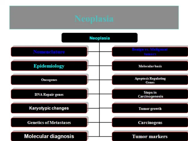
MOLECULAR BASIS OF CANCER
Three classes of normal regulatory genes principal targets of genetic damage. 1. Growth-promoting protooncogenes, 2. Growth-inhibiting cancer-suppressor genes (antioncogenes), 3. Genes that regulate programmed cell death, or apoptosis 4. DNA repair genes Mutant alleles of protooncogenes - oncogenes are considered dominant because they transform cells despite the presence of their normal counterpart. Tumor-suppressor genes both normal alleles of the must be damaged for transformation to occur - recessive oncogenes. Genes that regulate apoptosis may be dominant, as are protooncogenes, or may behave as cancer-suppressor genes.
Genetic changes that fuel tumor progression involved Growth-regulatory genes Genes regulate angiogenesis, invasion, and metastases Cancer related genes in the context of six fundamental changes in cell physiology that together dictate malignant phenotype 1. self-sufficiency 2. insensitivity to growth-inhibitory signals 3. Evasion of apoptosis 4. limitless replicative potential – overcome cellular senescence 5. sustained angiogenesis 6. ability to invade and metastasize
Carcinogenesis molecular basis of cancer Multistep process at both the phenotypic and the genetic levels A malignant neoplasm has several phenotypic attributes these characteristics are acquired in a stepwise fashion Tumor Progression. as excessive growth, local invasiveness, ability to form distant metastases. At the molecular level, progression results from accumulation of genetic lesions that in some instances are favored by defects in DNA repair.
A. Self-sufficiency in growth signal Oncogenes – mutant allele of protooncogenes genes that promote autonomous cell growth in cancer cells promote cell growth in the absence of normal growth-promoting signals products – oncoproteins resemble the product of normal protooncogenes except it is devoid of important regulatory elements and their production in transformed cells does not depend on growth factors or other external signals
Steps of cell proliferation • Binding of a GF to its specific receptor on CM • Transient and limited activation of the growth factor receptor, which in turn activates several signal-transducing proteins on the inner leaflets of the CM • Transmission of the transduced signal across the cytosol to the nucleus via a second messengers • Induction and activation of nuclear regulatory factors that initiate DNA transcription • Entry and progression of the cell into the cell cycle, resulting ultimately in cell division
Signal transduction cascade and Cell Cycle Regulation I. GROWTH FACTORS normal cell require stimulation by growth factors to undergo proliferation – most soluble GF are made by one cell type and act on a neighboring cell to stimulate proliferation ( paracrine action) Tumor cells 1. ability to synthesize the growth factor to which they responsive – autocrine 1.1 altered/mutated growth factor genes – excessive production Growth Factor 1.2 Most common Growth factor genes itself is not altered or mutated, but the products of other oncogene cause overexpression of growth factor genes cells are forced to secret large amount of Growth Factors
II. GROWTH FACTOR RECEPTORS 1. Mutation oncogenes encode growth factor receptors – overexpression of mutant receptor proteins deliver continuous mitogenic signals to cells even in the absence of the growth factors in the environment 2. Most common growth factor receptor genes itself is not altered or mutated but there are overexpression of GF from other sources – can render cancer cells hyperresponsive to normal levels of growth factors, a level that would not normally trigger proliferation HER2 (EGF receptor family ERBB1, ERBB2)in breast CA III.SIGNAL TRANSDUCING PROTEINS mutation in genes that encode various components of the signaling pathways ( overexpression of signaling proteins ) – couple with GF receptors to their nuclear targets RAS and ABL 30% of all human tumor contain mutated version of the RAS genes – undergo point mutation
Mutated RAS Genes produced Mutant RAS Proteins can bind GAPs but their GTPase activity fails to be augmented – mutant RAS is trapped in its activated GTP-bound form and the cell is led to believed that it must continue to proliferate Neurofibromin1
Nonreceptor-associated tyrosine kinase - signal transduction pathways ABL – protooncogene is dampened by negative regulatory domains chromosome 9 and is translocated to chromosome 22, where it fuses with part of the break point cluster region ( BCR) gene – BCR-ABL hybrid gene has a potent tyrosine kinase activity, and it activates several pathways including RAS-RAF cascade chronic myeloid leukemia certain acute leukemia IV. NUCLEAR TRANSCRIPTION FACTORS responder genes in the nucleus that orchestrate the cells orderly advance through the mitotic cycle – genes that regulate transcription of DNA oncogenes - MYC, MYB, JUN, FOS productions of oncoproteins MYC oncogene – associated with persistent expression or overexpression – overexpression of MYC oncoproteins
MYC proteins – bind to the DNA – transcriptional activation of several growth-related genes, including CYCLIN-DEPENDENT KINASE – drives cells into cell division translocation t(8:14) Burkitt’s lymphoma amplification breast, colon, lung and others Cyclin and Cyclin-dependent kinase growth promoting stimuli – entry of quiescent cells into cell cycle cyclin are synthesized during specific phase of the cell cycle and their function is to activate the CDK which is in inactive form and ones activated by cyclins phosphorylate crucial target proteins on completion of their task cyclin degenerate CDK inhibitors silence the CDK and exert negative control over the cell cycle
B.INSENSITIVITY TO GROWTH-INHIBITORY SIGNALS Normal cell antigrowth signal may cause dividing cell to go to G0 ( queiscence) or enter postmitotic, differentiated pool and lose replicative potential exert their effects on G1 to S phase check point of the cell cycle – this transition is controlled by RB protein 1.RB gene suppressor gene – product is a DNA-binding proteins RB protein serves as a brake in the advancement of cells from G1 to S phase when the cells are activated by growth factor – RB protein is inactivated by phosphorylation, ( phosphorylated RB – allow cell proliferation ) the break is released and the cells traverse the G1 to S phase when the cells enter S phase they are committed to divide without additional GF stimulation. During M phase the phosphate groups are removed from RB by cellular phosphate generating the dephosphorylated form of RB – prevent cell proliferation Mutated RB genes – absence or RB protein or its ability to sequester transcription factors is derailed by mutation