Download
1 / 27
270 likes | 388 Views
43y/o male pt heavey smoker 3 packs/D present to OPD c/o localized pain at left posterior side of the chest wall for 4 months duration associated with chronic non-productive cough. No Hx of trauma. PAST Hx: unremarkable.

E N D
43y/o male pt heavey smoker 3 packs/D present to OPD c/o localized pain at left posterior side of the chest wall for 4 months duration associated with chronic non-productive cough. • No Hx of trauma. • PAST Hx: unremarkable.
Ex: localized tenderness at left 9th rib (7cm distanant from posterior midline) + irregularity . • Chest Ex: bilateral EAE. • Routine blood works:within normal
Routine blood works:within normal. • CXR: unremarkable.
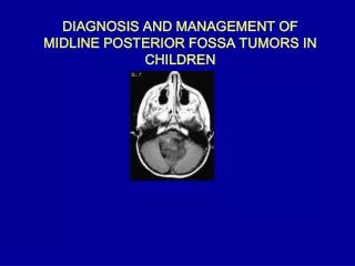
Pt reffered with MRI THORACIC SPINE (PRE AND POST CONTRAST) DONE ON 17-1-09: • SKIN MARKER WAS PLACED OVER THE TENDER AREA. • left posterior 9th rib abnormal enhancement lesion eroding the cortices (ant.&post.) with soft tissue component. • Right 8th intercostal space enhancing soft lesion (serpentine) with interforaminal extension.
CT SAN OF THE DORSAL SPINE AND RIBS (WITHOUT IV CONTRAST) 4-2-09: • Eroding and enhancing soft tissue lesion related to the posterior aspect of the left 9th rib with small extrapleural soft tissue mass. • It erodes the ant. And post. Aspect of the cortex with intramedullary extension. • Bilateral multiple small lung nodules w/w-out cavitation mainly upper lobes.
Differential diagnosis: • 1-LCH (lung and systemic disease). • 2-squamous cell ca :-lung +rib mets. • -head&neck+mets. • 3-chronic osteomyelitis(e,g. T.B. actinomyces). • 4-osteosarcoma +lung mets.(cavitation sometimes).
CT chest guided FNA CYTOLOGY done on7-2-09: SHOWED AGGREGATES OF HISTIOCYTES AND NEUTROPHILS.
Decision was made to do left 9th rib lesion excision and open lung Bx to reach Dx.
On the same day of O.R. 15-2-09: • CT- guided marking of lesion of 9th rib. • left 9th rib lesion excision. • through this window lung Bx was taken by thoracoscope. • CHEST TUBE WAS inserted IN LEFT SIDE AND WAS REMOVED 5 DAYS POST-OP.
Histiopatholgy report came as: (eosinophilic granuloma) histiocytosis X of both biopsies.
pt referred to hematologist which advice the pt to go to KFSH to take the chemothrrapy.
Alternative Names • Histiocytosis X; Langerhans cell histiocytosis; Eosinophilic granuloma; Pulmonary histiocytosis X ; Pulmonary Langerhans cell granulomatosis. • Histiocytosis is a general name for a group of syndromes that involve an abnormal increase in the number of immune cells called histiocytes.
There are three major classes of histiocytoses: • Langerhans cell histiocytosis, which is also called histiocytosis X. • Malignant histiocytosis syndrome (now known as T-cell lymphoma). • Non-Langerhans cell histiocytosis (also known as hemophagocytic syndrome). • We are going to focus only on Langerhans cell histiocytosis (histiocytosis X).
Eosinophilic Granuloma(Histiocytosis X): • uncommon interstitial lung disease. • The cause is unknown. • epidemiologically related to tobacco smoking. • ?autoimmune phenomenon with possible genetic and environmental predisposition. • young adults, primarily occurring in the third or fourth decades of life.
Pathophysiology: • PLCH is histologically characterized by abnormal infiltration of the lungs by activated Langerhans cells. • Langerhans cells are differentiated cells of the dendritic cell system and are closely related to the monocyte-macrophage line. • These antigen-presenting cells are normally found in the skin, reticuloendothelial system, heart, pleura, and lungs.
They may be identified by immunohistochemical staining or by the presence of Birbeck granules via electron microscopy. • usually manifests in a single organ; the lung. • About 4-20% of patients with PLCH also have cystic lesions in the bones. • Other organ systems are only rarely affected. • The accumulation found in the lungs is hypothesized to occur in response to exposure to cigarette smoke.
Frequency: • a rare disorder and the true prevalence is unknown. • less than 5% of patients who underwent lung biopsy for the diagnosis of interstitial lung disease. • affect roughly 1 in 200,000 people each year.
Race: • Because of the rarity of the disease, no definitive epidemiologic data related to racial background are available. • Sex: • No sex predilection is recognized. • Age: • The peak incidence occurs in the 20- to 40-year age bracket.
Symptoms: • variable. • 25% of patients are asymptomatic • incidental findings on chest radiographs • 75%present with respiratory or constitutional symptoms. • Nonproductive cough (56-70%) • Dyspnea (40%) • Fatigue (30%) • Weight loss (20-30%) • Chest pain (21%) • Spontaneous pneumothorax, which may be recurrent, is a classic presentation found in 10-20% of patients. • Fever (15%) • Cystic bone lesions (4-20%): These may be painful and may predispose the patient to pathologic fracture.
Physical Ex: • Patients with PLCH present with nonspecific physical findings. • Laboratory Studies: • Results are nonspecific. • Peripheral eosinophilia is not observed.
Imaging Studies: • Chest radiographs characteristically reveal bilateral, symmetric, ill-defined nodules and reticulonodular infiltrates. • As the disease progresses, cystic lesions appear. • An upper-zone predominance of radiographic findings with sparing of the costophrenic angles is typically observed.
CT chest: • Pathognomonic findings (nodules and cysts, predominantly in the mid and upper lung zones, with sparing of the costophrenic regions. • The nodules may be cavitary and variable in size.
Open or thoracoscopic lung biopsy is the most sensitive and specific diagnostic modality.
Histologic Findings: • granulomatous nodules composed of Langerhans cells as well as eosinophils, macrophages, lymphocytes, plasma cells, and fibroblasts. • Electron microscopy helps in identifying Langerhans cells by demonstrating the presence of diagnostic pentilaminar cytoplasmic inclusion bodies, or Birbeck granules (x-bodies). • As the disease progresses, cavitation occurs as a result of this destruction. • The nodule fibroses, eventually forming a stellate scar.
Management: • Smoking cessation is the most important medical intervention. • Smoking cessation often stabilizes the disease and sometimes leads to regression. • corticosteroids is controversial. • Chemotherapeutic agents. • Lung transplantation is an option for select patients with advanced disease. • Radiation therapy or surgery may also be used to treat bone lesions. • Breathing support (with a breathing machine) • Physical therapy.
Complications: • Spontaneous pneumothorax (10-20%). • increased risk of malignancy, including Hodgkin and non-Hodgkin lymphoma, myeloproliferative disorders, and bronchogenic carcinoma. • Pathologic fracture may occur at the site of bone lesions. • Interstitial Pulmonary fibrosis. • Pulmonary artery hypertension and cor pulmonale may develop as a result of hypoxemia and/or vascular disruption due to PLCH lesions.
Prognosis: • Prognosis varies and is related to smoking cessation. • Most patients who continue to smoke experience disease progression, but for those who successfully quit smoking, the disease often stabilizes regresses.