Download

1 / 52
520 likes | 665 Views
CASE STUDY FOR M-1 STUDENTS. CASE #1 Patient presents to his doctor with complaint of back pain with increasing intensity. L-2. Diagnosis: 1-Degenerative Disc disease at (L2-3 and L4-5) . L-5. 1 1. Add red arrows & captions that

E N D
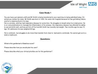
CASE #1 Patient presents to his doctor with complaint of back pain with increasing intensity. L-2 Diagnosis: 1-Degenerative Disc disease at (L2-3 and L4-5) L-5 11
Add red arrows & captions that confirm the diagnosis and /or other abnormalities. Use blue arrows to indicate 3 normal structures . Your interpretation here. RADIOLOGY EXAM: Lateral Lumbar Spine CLINICAL INDICATION: Back pain increasing in intensity. REPORT: The patient has degeneration of IV discs at the L2- L3 level and the L4-L5 level . CONCLUSION: Patient shows signs of degenerative disc disease affecting L2-3 and L4-5. Normal L2 vertebrae L-2 Disc degeneration at L2-L3 with bone spurs. L4-5 foramen Normal L4 vertebrae L-5 Disc degeneration at L4-L5 1 1
Three bullet points about pathology identified • OR • Management of the identified process • 50 words or less • Source of pain is due to inflammation and abnormal micromotion instability. • Many cases can be managed by anti-inflammatory medication, physical therapy and bed rest. • Most common cause of disc degeneration is aging. 1 1
SPINE CASE #2 11 year old male in the trauma room following a head on collision. Diagnosis: Atlanto-occipital dislocation 2
Add red arrows & captions that confirm the diagnosis and /or other abnormalities. Use blue arrows to indicate 3 normal structures . RADIOLOGY EXAM: Cross table lateral cervical spine x-ray. CLINICAL HISTORY: 11 yom in trauma room following a head on collision REPORT: The space between the skull and C1 is widened greater than normal. The occipital condyles are not resting on the superior articular surfaces of C-1. CONCLUSION: The high impact of the collision caused disruption of the Atlanto-occipital joint. Cross table lateral Abnormal curvature of C-Spine Widened C-1 occipital space Normal C-5 vert. body Normal IV foramen Normal spinous process 2
Three bullet points about pathology identified. • OR • Management of the identified process • 50 words or less • The membranes and ligaments holding the skull onto C1 are damaged during this type of injury causing the skull to dislocate from the rest of the spine. • An Atlanto-occipital dislocation may occur without a fracture of the fracture of the C1 vertebra. • Atlanto-occipital dislocations can often be fatal, even without a fracture of the c1 vertebra. 2
CASE # 3 Patient goes to the doctor with the complaint of pain and reduced range of motion of his back. Diagnosis: Ankylosing spondylitis “Bamboo Spine” 3
Add red arrows & captions that confirm the diagnosis and /or other abnormalities. Use blue arrows to indicate 3 normal structures . RADIOLOGY EXAM: AP and Lateral Lumbar Spine CLINICAL INDICATION: Decresed range of motion. Chronic lower back pain. REPORT: Fused vertebral bodies at multiple disc levels. Scoliosis in the lower thoracic region. Fused sacroiliac joints. CONCLUSION: The fused vertebral bodies and fused sacroiliac joints indicated that the diagnosis is Ankylosing spondylitis. Normal IV disc Scoliosis Normal IV foramen Fused vertebral bodies Sacrum 3
Three bullet points about the pathology OR • Management of the identified process • 50 words or less • Ankylosing spondylitis is a form of arthritis, primarily affection the spine • Most people with AS have a gene that produces the genetic marker for the protein HLA-B27 • Can also cause swelling in other areas, such as shoulders, hips, ribs, heels and small joints of the hands and feet 3
20 YEAR OLD MALE WITH BACK PAIN Diagnosis: Spondylolysis of L-5 4
Add red arrows & captions that confirm the diagnosis and /or other abnormalities. Use blue arrows to indicate 3 normal structures . Rib RADIOLOGY EXAM: Lateral Lumbar spine x-ray CLINICAL INDICATION: Lower back pain REPORT: X-ray shows defect indicating a fracture at the L5 inferior articular process at the pars interarticularis. The other zygopophysial joints appear normal. CONCLUSION: Spondylolysis of the L5 vertebra Normal L2 vertebral body Intervertebral foramen Pars defect. 4
Three bullet points about pathology identified OR Management of the identified process 50 words or less May be caused by failure of the Centrum of L5 to unite adequately with the neural arches at the neurocentral joint during development. Bracing , Rest and physical therapy are used for the management of pain 4
Patient has back pain and positive lab work up for proteinuria. Diagnosis: Multiple myeloma 5
Add red arrows & captions that confirm the diagnosis and /or other abnormalities. Use blue arrows to indicate 3 normal structures . RADIOLOGY EXAM: Lateral lumbar spine CLINICAL INDICATION: Back pain and positive labs for proteinuria. REPORT: Osteoporotic vertebral bodies in the lumbar spine with bowed endplates. CONCLUSION: Due to positive lab workup for proteinuria and areas of osteoporosis in the spine and vertebra, multiple myeloma has to be considered. Flattened vertebra Osteoporotic bone Foramen Normal bowel gas Spinous process Endplate bowing 5
Three bullet points about pathology identified • OR • Management of the identified process • Multiple Myeloma begins when plasma cells become abnormal and continue to divide. • Over time cells collect in bone marrow crowding normal blood cells and causing extensive destruction to bone leading to osteoporosis. • Abnormal plasma cells secrete abnormal; proteins which can lead to clotting and kidney failure. 5
80 year old woman goes to the doctor with pain in her neck. She is currently being treated For Rheumatoid arthritis Diagnosis; C 1-2 Subluxation 6
Add red arrows & captions that confirm the diagnosis and /or other abnormalities. Use blue arrows to indicate 3 normal structures . RADIOLOGY EXAM: Lateral C-spine X-ray CLINICAL INDICATION: 80 YOF rheumatoid arthritis patient with neck pain. REPORT: The distance between the posterior surface of the anterior tubercle of C-1 and the anterior surface of the dens is markedly increased indicating that there is disruption of the transverse ligament of C1 and C2. There is also degenerative disc narrowing at C3 through C6. CONCLUSION:C1-C2 Subluxation Degenerative disc narrowing at C3-C6 Note the position of the posterior tubercle of C-1 Increased distance between the anterior arch of C1 and the dens Normal C-6 spinous process Normal C7 vertebral body Normal C7-T1 disc space 6
Three bullet points about pathology identified • OR • Management of the identified process • 50 words or less • C1-C2 subluxation can cause pain in flexion because C1 will compress the spinal cord. • Rheumatoid arthritis can cause stretching and destruction of the transverse ligament which allows C1 to move forward relative to C2. • C1-C2 subluxation tends to occur because of pannus formation at the gliding synovial joints. 6
Case #7 A 75 year old woman goes to the ED complaining of neck pain. She tells doctor that she fell down her steps(4) yesterday. Study the image—add your diagnosis in the place provided and send back to Penelope.Al-Emam@uscmed.sc.edu Diagnosis: Fracture of C-2 (Odontoid process)
Add red arrows & captions that confirm the diagnosis and /or other abnormalities. Use blue arrows to indicate 3 normal structures . RADIOLOGY EXAM: Lateral C-Spine X-Ray CLINICAL INDICATION: Pain in neck after falling down steps. REPORT: There appears to be a transverse fracture at the base of the dens with subsequent anterior shift of the C1 vertebra and the skull. The anterior shift in the C1 vertebra indicates a possible impingement of the spinal cord. Degenerative disc narrowing is also noted at C3-4, C4-5 mad C5-6 CONCLUSION: There is a Type ll Fracture of the C2 Odontoid process causing and anterior shift of the C1 vertebra indicates a possible impingement of the spinal canal. Fracture of the odontoid Posterior tubercle of Atlas Vertebral body of C6 IV disc space
Three bullet points about pathology identified • OR • Management of the identified process • 50 words or less • Non-operative means are contraindicated due to the unstable fracture. • Type II fracture indicated internal fixation as the primary management. • A fusion of the C1 and C2 vertebrae is created using wire and midline bone grafting.
70 year old male is experiencing pain & lower extremity paralysis Diagnosis: Multiple Metastatic Lesions 8
Add red arrows & captions that confirm the diagnosis and /or other abnormalities. Use blue arrows to indicate 3 normal structures . RADIOLOGY EXAM: CT of Thoracic spine CLINICAL INDICATION: Back pain and lower extremity paralysis REPORT: Red arrows show area of destructive lesions protruding posteriorly from the vertebral canal to the spinous processes. CONCLUSION: Multiple metastatic lesions are impinging on the nerves and spinal cord which leads to lower extremity paralysis and pain experienced by the patient. 8
Three bullet points about pathology identified OR Management of the identified process 50 words or less Management of metastatic lesions could include either radiation therapy, removal of the tumors through open surgery or Percutaneous Inage-Guided Vertebral body Augmentation. Radiation and open surgery are the two most common treatments. Radiation offers a non-invasive, less immediate risk option. Percutaneous Image-Guided Vertebral Body Augmentation uses bone cement to relieve pain from spinal tumors and stabilizes the spine. It is considered less-invasive that open surgery. Vertebral body Spinous process of Thoracic vertebra Rib 8
25 year old male with neck pain following MVA Diagnosis: C5 fracture 9
Add red arrows & captions that confirm the diagnosis and /or other abnormalities. Use blue arrows to indicate 3 normal structures . RADIOLOGY EXAM: Lateral C-Spine x-ray CLINICAL INDICATION: Neck pain following MVA REPORT: Fracture of the anterior portion of the C5 vertebral body. There is widening of facet joints and interspinous spaces of the C5 cervical vertebra. CONCLUSION: C5 fracture Spinous process Aligned facet joint Fragmented anterior portion of vertebral body Widened facet joint Trachea 9
Three bullet points about pathology identified • OR • Management of the identified process • 50 words or less • Fractures of this type are loosely referred to as “tear-drop” fractures based of appearance of the triangular displaced vertebral body portion. • Anterior column trauma may be result of axial loading injuries including a combination of extreme compression, extension, flexion, or rotational events. • Often, anterior spinal injuries accompany loss of motor function, temperature sensation, pain but maintaining proprioception. 9
SPINE CASE #10 Patient goes to the doctor with Complaint of back pain. Please Study the images-add Your diagnosis and return to me @ Penelope.Al-Emam@uscmed.sc.edu Diagnosis--Kypho-scoliosis 10
Add red arrows & captions that confirm the diagnosis and /or other abnormalities. Use blue arrows to indicate 3 normal structures . RADIOLOGY EXAM: AP & Lateral Chest. CLINICAL INDICATION: Back pain REPORT: Ap x-ray shows curvature of the thoracic spine indicative of scoliosis concave to the left at T-9. Lateral x-ray shows an exaggerated primary kyphotic curv of the thoracic spine. CONCLUSION: Kyphotic scoliosis Clavicle Scoliosis and kyphosis of the thoracic spine Diaphragm IV disk space 10
Add red arrows & captions that confirm the diagnosis and /or other abnormalities. Use blue arrows to indicate 3 normal structures . Three bullet points about pathology identified OR Management of the identified process 50 words or less Patient can undergo surgery for spinal decompression and spinal fusion with brackets and screws. 10
33 year old truck driver goes to the Health center for his required DOT physical as a new employee. Diagnosis: Bifid spinous processes C-7, T-1 & T-2 11
Add red arrows & captions that confirm the diagnosis and /or other abnormalities. Use blue arrows to indicate 3 normal structures . RADIOLOGY EXAM: Frontal Chest X-ray CLINICAL INDICATION: Required Physical REPORT: Spina Bifida Occulta in Spinous Processes of C7, T1, and T2 CONCLUSION: see report INTERPRETER’S NAME_______________ DATE______________ Clavicle Bifid spinous processes Normal spinal process Lt. 4th rib 11
Three bullet points about pathology identified • OR • Management of the identified process • 50 words or less • Spina bifida occulta occurs when lamina do not merge during development, thus creating a bifid spinous process. • Twenty –five percent of the population has this abnormality, but it is generally asymptomatic. • The meninges and spinal cord are generally not affected due to this defect alone. 11
A 50 year old man goes to his general physician with the complaint of a sore throat x 2 weeks.--- A soft tissue lateral neck x-ray was done. Diagnosis: Segmentation anomaly of C5/6 12
Add red arrows & captions that confirm the diagnosis and /or other abnormalities. Use blue arrows to indicate 3 normal structures . RADIOLOGY EXAM: Soft tissue lateral neck CLINICAL INDICATION: Sore throat x 2 weeks REPORT: Fusion of vertebral body C5-6 with bony bridge across the disc space. Transverse and spinous processes appear normal and healthy. No visible fractures, possible edema of the anterior soft tissue at level of C5-C6, no apparent IV disk between C5-C6 mainly bone present CONCLUSION: Segmentation Anomaly of C5-C6 C2 Spinous Process C3 vertebral body Hyoid bone Fused vertebral bodies 12
Three bullet points about pathology identified • OR • Management of the identified process • 50 words or less. • Congenital • Defective segmentation of the developing tissue within the spine • Administer anti-inflammatory medication to help with sore throat. 12
50 year old man falls from a ladder CT scans done in the emergency room. Diagnosis: Compression fracture of T-12 13