Download
1 / 18
180 likes | 276 Views
Use the simplest technology that will answer the scientific question!. Fluorometer Low light digital imaging Confocal microscopy Two photon microscopy. Use the simplest technology that will answer the scientific question!. Population response adequate cell/cell heterogeneity not crucial

E N D
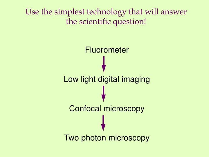
Use the simplest technology that will answer the scientific question! Fluorometer Low light digital imaging Confocal microscopy Two photon microscopy
Use the simplest technology that will answer the scientific question! Population response adequate cell/cell heterogeneity not crucial Cell:cell heterogeneity important intracellular heterogeneity not crucial Samples up to 50 µm thick, cell:cell or subcellular resolution needed Thick specimens up to several hundred µm thick, cell:cell or subcellular resolution needed
Selecting a system The decision points speed depth of focus resolution sensitivity
Selecting a system • The cleanest triage point: speed • If you need detailed image information at greater than 3 frames/second, think about the Perkin-Elmer/Solamere fast scanner. • But first ask… • Can you do your experiment with a subset of the image? Maybe you can use other systems… • Do you need simultaneous dual or triple emissions? Maybe you should use other systems… • Are you just thinking you MAY need to go fast, but for 90% of what you do another system is of better use?
Selecting a system • The next cleanest triage point: depth • If you need to image into specimens thicker than 60 m, think about multi-photon microscopy. • But first ask… • Do you know if you can get your reporter molecule in that deep? • Could you just look at the sample from the other side as well? • Does your tissue happen to be a really great confocal sample or a really lousy two-photon sample?
Selecting a system • The lousiest triage points: resolution and sensitivity • Extremely dependent on how you set up your experiment. Personal rather than absolute. Often comes down to available options or sample preparation. Fortunately, simple tests to see if any given microscope gives adequate results STRATEGY: Ask if the device can give good results with YOUR sample that YOU want to study, not some test specimen that is completely irrelevant.
Selecting a system Get the sample that everyone hates. The one that photobleaches before you can even find the cell you are interested in. fixed: FITC without antifades (antioxidants) live: BCECF 1. Set your detectors as sensitive as possible. 2. Set your light source as low as possible. 3. Increase light and/or average the noisy image until you get an image that you can live with (compulsive people can estimate Signal/Bkground noise ratios) 4. Collect a series of ~30 images at 1/sec, then at 1/10 second Define sensitivity by ability to minimize cell damage
Selecting a system Define sensitivity by ability to minimize cell damage Cell brightness time IF percent fluorescence loss is a constant with each time point, then it is due to photobleaching
Selecting a system Define sensitivity by ability to minimize cell damage Cell brightness time IF fluorescence loss does not change as a function of sampling rate, then it is due to dye leakage/diffusion with no evident photobleaching. This is a more sensitive system, able to image without damage.
Selecting a system • Invite vendors in for extended demos • Replace color glossy brochures with your own experience • Get your hands involved • Don’t let them run the device all the time • You need to see how user-friendly it is • Make sure you understand the features and capabilities Even better, try them out to see if they work • Work with lousy samples • Dim • One label is massively brighter than another • Bad pairing of colors with lots of bleed over
Funding a system NSF: Multi-User Equipment and Instrumentation Resources Grant (MUE) first Monday in October up to $400,000 minimum $40,000 instrument (30% cost share required) no “imaging facility” bundle of instruments goal: further NSF-funded, or NSF applicable research with SOATs need minimum of 3 independent investigators “Some” users should have active NSF funding
Funding a system NIH: Shared Instrumentation Grant (S10) once a year deadline, March up to $500,000 minimum $100,000 instrument (cost share) no “imaging facility” bundle of instruments goal: further NIH-funded research with SOATs need minimum of 3 NIH-funded investigators suggest 4-6 in user group, not 16 Biological question not under fire feasibility and sensibility of equipment use under review preliminary data crucial to demonstrate experience and feasibility
Funding a system NIH: Shared Instrumentation Grant (S10) grant pieces important: read the instructions User group and instrument should be a tight fit. requested options should be used by 3 or more investigators justify everything: no justification, no lens make it clear how the instrument… improves your efficiency and progress gets around existing bottlenecks (time, availability, capability) provides new areas for exploration Make sure the expertise is available to help users technician should be trained optical expertise should be evident Place in a core facility and outline how sharing works leave time for outside users captive instruments tick off the reviewers
How to get preliminary data • NIH/NCRR Biomedical Technology Resources • http://www.ncrr.nih.gov / ncrrprog / btdir / btdirectory.asp • Scientists at these centers ensure that biomedical investigators who have NIH-supported projects may gain access to the newest and most advanced technologies, techniques, and methodologies. • Laser Applications • Center for Fluorescence Spectroscopy (Baltimore, MD) • Laboratory for Fluorescence Dynamics (Urbana, IL) ** • Laser Biomedical Research Center (Cambridge, MA) • Laser Microbeam and Medical Program (Irvine, CA)** • National Flow Cytometry and Sorting Research Resource (Los Alamos, NM) • Ultrafast Optical Processes Laboratory (Philadelphia, PA)** • Microscopy • National Center for Microscopy and Imaging Research (La Jolla, CA) ** • National Resource for Automated Molecular Microscopy (La Jolla, CA) • Resource for the Visualization of Biological Complexity (Albany, NY) • Three-Dimensional Electron Microscopy of Cells (Boulder, CO) • Three-Dimensional Electron Microscopy of Macromolecules (Houston, TX)
Establishing a microscopy facility • Prepare the space • Power • Cooling • Air flow • Wet lab/surgery facilities • Prepare the skills and peripheral tools • Training staff and ongoing user training • Sign up strategies for equitable sharing • Off-line analyses • Prepare for disaster • Fiscal survival • Replacements/repair • Penalties
Post-delivery depression • Living with the system after purchase • Monitoring performance/ Standardize response • “..but I’m sure it was more sensitive last week” • Fixed hour for PM/cleanup weekly • Living with the users after purchase • Data archiving • Get users off the microscope for data analysis and printing images • “Tell me when it’s broken or I can’t fix it”