Download
1 / 39
390 likes | 400 Views
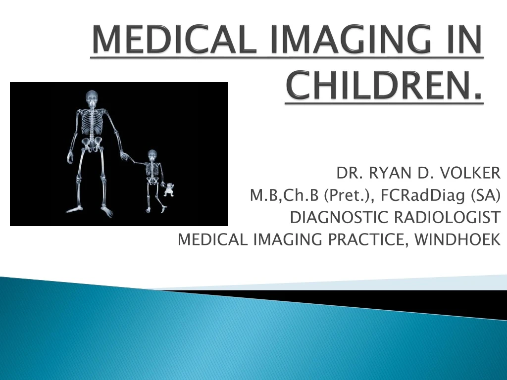
MEDICAL IMAGING IN CHILDREN. DR. RYAN D. VOLKER M.B,Ch.B (Pret.), FCRadDiag (SA) DIAGNOSTIC RADIOLOGIST MEDICAL IMAGING PRACTICE, WINDHOEK. MAXIMISING POSITIVE FINDINGS WHILE CURBING THE COSTS, i.e “GETTING MORE BANG FOR YOUR BUCK!”. The following still holds true:.

E N D
MEDICAL IMAGING IN CHILDREN. DR. RYAN D. VOLKER M.B,Ch.B (Pret.), FCRadDiag (SA) DIAGNOSTIC RADIOLOGIST MEDICAL IMAGING PRACTICE, WINDHOEK
MAXIMISING POSITIVE FINDINGS WHILE CURBING THE COSTS, i.e “GETTING MORE BANG FOR YOUR BUCK!”
The following still holds true: • Children are not just and can never just be regarded as small adults in terms of disease profile, management or IMAGING.
IMAGING MODALITIES AVAILABLE: • Plain X-Rays • Screening investigations, e.g barium studies • Ultrasound • Computed Tomography – CT scan • Magnetic Resonance Imaging - MRI
UNIQUE ISSUES PERTAINING: • COST OF MODALITY VS. BENEFIT: • X-Rays are most cost-effective > ultrasound >>CT (sedation) >>> MRI ( ++ sedation ). • Much information to be gained on X-rays and ultrasound vs adults. • RADIATION DOSE VS. BENEFIT: • Children are ++ radiation-sensitive. • Ultrasound, MRI no radiation vs high doses with CT >> X-rays. • CT dose up to 100x normal X-ray. ORDER THE CORRECT X-RAY ORDER ONLY NECESSARY X-RAYS
TODAY’S DISCUSSION: • We will concentrate on the cost-effective and relatively safe modalities that are easily accessible: • Plain X-Rays. • Screening investigations. • Ultrasound.
PLAIN X-RAYS THAT ARE OF LITTLE VALUE: • Skull X-ray and facial bones. • Significant intracranial injuries may be present with a normal skull X-ray. • Facial bone fractures often missed on plain X-rays. • X-rays of the paranasal sinuses. • Particularly in children under six years. • Very limited anatomy visualised, e.g OMU.
THE CHEST X-RAY: • Cost-effective and easily accessible • Relatively low-dose modality • Provides important information quickly in neonates and children • Confirm, help with diagnosis • Can guide / change management approach • Useful in monitoring therapeutic outcomes
THE NEONATE: • Premature baby – Hyaline membrane disease. • Baby delivered by Caesarean section – transient tachypnoea / wet lung. • Meconium aspiration syndrome.
INFANTS AND CHILDREN: • Bronchiolitis caused by the Respiratory Syncytial Virus. • Bronchopneumonia usually in older children – bacterial infection and PTB.
THE ABDOMINAL X-RAY: • Neonate: • Prematurity and ICU – necrotizing enterocolitis. • Congenital conditions – duodenal atresia. • Acquired conditions – intussusception. • Infants and children: • Abdominal pain due to faecal loading. • Appendicitis – better diagnosed on ultrasound.
DUODENAL ATRESIA DOUBLE-BUBBLE SIGN
INTUSSUSCEPTION 2-3 year old infant Red-currant stool
ACUTE APPENDICITIS Sentinel loops Blind-ending tubular swollen appendix
X-RAYS OF THE LIMBS: • Injuries: • Non-accidental injuries – important to recognise. • Accidental fractures, e.g greenstick fractures. • Metabolic conditions: • Congenital • Rickets
ACCIDENTAL FRACTURES Common greenstick wrist fractures Supracondylar fractures
RICKETS Rachitic Rosary Metaphyseal fraying, splaying and cupping
SCREENING STUDIES: • Barium meal – commonly needed for the diagnosis of suspected reflux. Also performed to assess for malrotation and midgutvolvulus. • Barium enema – most often requested to exclude Hirschsprung’s disease. • Voiding cysto-urethrogram – for children with repeated UTI’s, especially boys. Most often to exclude posterior urethral valves, VUR.
HIRSCHSPRUNG’S DISEASE Short segment – inverse recto-sigmoid ratio Long segment
VESICO-URETERIC REFLUX Grading of VUR Grade III VUR
ULTRASOUND: • Cost is slightly more than plain X-ray. • Completely safe with no radiation danger as sound waves are utilised. • Slightly less accessible than X-rays, particularly in rural areas. • Requires a degree of training in order to perform and interpret.
COMMON DIAGNOSES: • CRANIAL SONAR: Hydrocephalus, germinal matrix haemorrhage in premature babies. • CHEST SONAR: Pleural effusions. • ABDOMINAL SONAR: Hypertrophic pyloric stenosis, appendicitis, intussusception, mesenteric adenitis, hydronephrosis, paediatric neoplasms, e.gnephroblastoma, neuroblastoma, hepatoblastoma. • MUSCULOSKELETAL SONAR: Congenital hip dysplasia, injuries.
CRANIAL ULTRASOUND: Hydrocephalus Grade I germinal matrix haemorrhage
ABDOMINAL ULTRASOUND Hypertrophic pyloric stenosis Barium meal
ABDOMINAL ULTRASOUND Mesenteric adenitis
ABDOMINAL ULTRASOUND Severe hydronephrosis Nephroblastoma