Download
1 / 15
150 likes | 269 Views
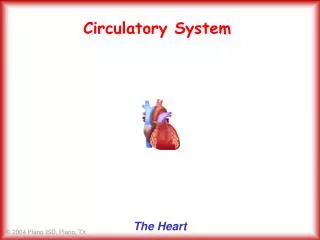
The Heart. Location . The adult heart is approx. the size of a closed fist. It resides in the thoracic cavity between the lungs in an area called the mediastinum. The top is rounded and called the base, the bottom has a point called the apex. . Anatomy of Heart. Three layers

E N D
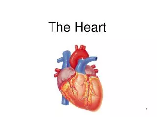
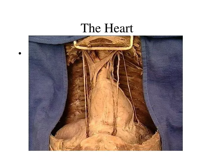
Location • The adult heart is approx. the size of a closed fist. It resides in the thoracic cavity between the lungs in an area called the mediastinum. The top is rounded and called the base, the bottom has a point called the apex.
Anatomy of Heart Three layers • Outermost is the pericardium – or pericardial sac: double layered – tough fibrous connective tissue • Visceral – on heart surface • Parietal – towards the cavity • Pericardial cavity – pericardial fluid - serous
Middle Layer – myocardium – muscular layer Muscle cells are connected via intercalated discs - carry conduction in a ‘domino effect’ Innermost layer – endocardium – epithelial layer – simple squamous epithelium over a tough connective tissue Valves are a result of the endocardium folding – valves between the atrium and ventricle = AV valve, atrioventricular valve or tricuspid [R] /bicuspid [L]. -valves between the ventricles and vessels = semilunar valves or pulmonic [R]/aortic[L] Chambers: right & left atria right & left ventricles Major Vessels: vena cava, aorta, pulmonary veins, pulmonary trunk
ElectroCardioGraph [EKG/ECG] • Electrical conduction of the heart can be monitored by electrodes on body surface. • Not a direct measure of mechanical events, force of contraction or blood pressure.
The normal EKG consists of a P wave, the QRS complex, and a T wave.
The P wave indicates that the atria (the two upper chambers of the heart) are contracting to pump out blood. the "QRS complex." This part indicates that the ventricles (the two lower chambers of the heart) are contracting to pump out blood to the body The ST segment indicates the amount of time from the end of the contraction of the ventricles to the beginning of the rest period before the ventricles begin to contract for the next beat. The T wave indicates the resting period of the ventricles.
arrhythmia An arrhythmia (also called dysrhythmia) is an abnormal rhythm of the heart, which can cause the heart to pump less effectively. Arrhythmias can cause problems with contractions of the heart chambers by: not allowing the chambers to fill with an adequate amount of blood, because an electrical signal is causing the heart to pump too fast. not allowing a sufficient amount of blood to be pumped out to the body, because an electrical signal is causing the heart to pump too slowly or too irregularly.