Download

1 / 17
170 likes | 321 Views
Table 1.1. Table 1.1. Table 1.1. Table 1.1. Table 1.1. Regional Terms. Two major divisions of body: Axial Head, neck, and trunk Appendicular Limbs Regional terms designate specific areas. Upper limb. Cephalic. Acromial. Frontal. Brachial (arm). Orbital. Antecubital. Nasal.

E N D
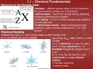
Regional Terms • Two major divisions of body: • Axial • Head, neck, and trunk • Appendicular • Limbs • Regional terms designate specific areas
Upper limb Cephalic Acromial Frontal Brachial (arm) Orbital Antecubital Nasal Antebrachial (forearm) Oral Mental Carpal (wrist) Cervical Manus (hand) Thoracic Palmar Axillary Pollex Mammary Digital Sternal Abdominal Lower limb Umbilical Coxal (hip) Pelvic Femoral (thigh) Inguinal (groin) Patellar Crural (leg) Fibular or peroneal Pubic (genital) Pedal (foot) Tarsal (ankle) Thorax Metatarsal Abdomen Digital Back (Dorsum) Hallux (a) Anterior/Ventral Figure 1.7a
Upper limb Cephalic Otic Acromial Occipital (back of head) Brachial (arm) Olecranal Cervical Antebrachial (forearm) Back (dorsal) Manus (hand) Scapular Metacarpal Vertebral Digital Lumbar Lower limb Sacral Femoral (thigh) Gluteal Popliteal Perineal (between anus and external genitalia) Sural (calf) Fibular or peroneal Pedal (foot) Thorax Abdomen Back (Dorsum) Calcaneal Plantar (b) Posterior/Dorsal Figure 1.7b
Frontal plane Median (midsagittal) plane Transverse plane (a) Frontal section (through torso) (b) Transverse section (through torso, inferior view) (c) Median section (midsagittal) Pancreas Aorta Spleen Liver Spinal cord Intestines Rectum Spleen Left and right lungs Liver Heart Body wall Vertebral column Stomach Arm Subcutaneous fat layer Figure 1.8
Cranial cavity Dorsal body cavity Ventral body cavity Cranial cavity (contains brain) Vertebral cavity Superior mediastinum Dorsal body cavity Thoracic cavity (contains heart and lungs) Pleural cavity Pericardial cavity within the mediastinum Vertebral cavity (contains spinal cord) Ventral body cavity (thoracic and abdominopelvic cavities) Diaphragm Abdominal cavity (contains digestive viscera) Abdomino- pelvic cavity Pelvic cavity (contains urinary bladder, reproductive organs, and rectum) (a) Lateral view (b) Anterior view Figure 1.9a-b
Serous Membrane (Serosa) • Thin, double-layered membrane separated by serous fluid • Parietal serosa lines internal body walls • Visceral serosa covers the internal organs
Outer balloon wall (comparable to parietal serosa) Air (comparable to serous cavity) Inner balloon wall (comparable to visceral serosa) Heart Parietal pericardium Pericardial space with serous fluid Visceral pericardium (b) The serosae associated with the heart. Figure 1.10a-b
Right upper quadrant (RUQ) Left upper quadrant (LUQ) Right lower quadrant (RLQ) Left lower quadrant (LLQ) Figure 1.11
Diaphragm Liver Right hypochondriac region Left hypochondriac region Epigastric region Stomach Gallbladder Transverse colon of large intestine Ascending colon of large intestine Right lumbar region Left lumbar region Umbilical region Descending colon of large intestine Small intestine Cecum Initial part of sigmoid colon Right iliac (inguinal) region Hypogastric (pubic) region Left iliac (inguinal) region Appendix Urinary bladder (a) Nine regions delineated by four planes (b) Anterior view of the nine regions showing the superficial organs Figure 1.12
Other Body Cavities • Oral and digestive cavities • Nasal cavity • Orbital cavities • Middle ear cavities • Synovial cavities