Download
1 / 15
170 likes | 328 Views
Calcium binding proteins. Maria Tereshina Vsevolod Belousov. Calcium binding protein CaBP2 Results of CD search by BLAST reveal two Efh domains. Calcium binding protein CaBP2 Results of CD search by BLAST reveal two Efh domains, each consisting of ef-hand motifs.

E N D
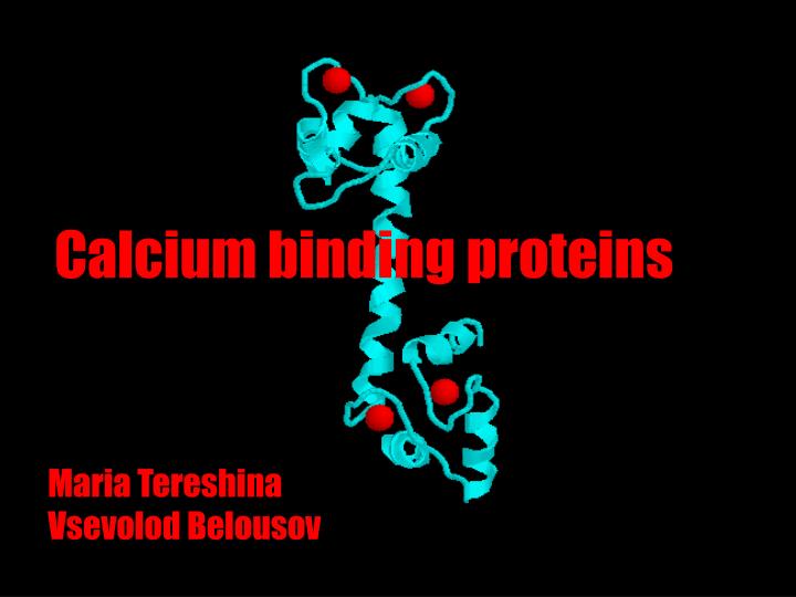
Calcium binding proteins Maria Tereshina Vsevolod Belousov
Calcium binding protein CaBP2 Results of CD search by BLAST reveal two Efh domains
Calcium binding protein CaBP2 Results of CD search by BLAST reveal two Efh domains, each consisting of ef-hand motifs
Stricture of typical ef-hand containing protein, calmodulin +4Ca2+
N-terminal domain of calmodulin with bound Ca2+ Yellow- key positions in ef-hand, coordinating Ca2+ (usually Asp & Glu)
N-terminal domain of calmodulin with bound Ca2+ N-terminal domain of calmodulin with bound Ca2+ Yellow- key positions in ef-hand, coordinating Ca2+ (usually Asp & Glu)
Calsequestrin- ER protein with negatively charged surface that binds Ca2+ with low affinity
Crystal structure of calsequestrin. Red spacefill- Asp & Glu residues
Structure of C2 domain from PKCa Yellow- Asp residues coordinating Ca2+
Structure of Ca2+-binding domain of collagenase Conformation changes upon Ca2+ load
Structure of Ca2+-binding domain of collagenase Conformation changes upon Ca2+ load
CONCLUSION The diversity of calcium binding proteins is great!