Download

1 / 88
960 likes | 1.74k Views
Acid-base disorders and interpretation of arterial blood gases. Juan F. Sanchez, MD Assistant Professor of Medicine, Pulmonary and Critical Care Medicine Texas A&M College of Medicine Scott and White Memorial Hospital. Objectives. Describe physiology involved in acid base balance of the body

E N D
Acid-base disorders and interpretation of arterial blood gases Juan F. Sanchez, MD Assistant Professor of Medicine, Pulmonary and Critical Care Medicine Texas A&M College of Medicine Scott and White Memorial Hospital
Objectives • Describe physiology involved in acid base balance of the body • Review causes and treatments of acid base disorders • Identify normal arterial blood gas values • Interpret results of ABG samples • Interpret oxygenation state of a patient using reported ABG values
Interpretation of ABG • Very important for health care providers • Usefulness of this tool depends on being able to interpret correctly the results • Critically ill patients
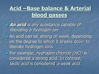
Acid base disorders • 12000 to 15000 mEq of volatile acids are produced daily by body and excreted as CO2 by lungs • 1 mEq / kg / day of non-volatile acids (sulfuric and phosphoric acids) are produced daily by body and excreted by the kidneys • The pH of body fluids is determined by the amount of acid produced, the buffering capacity and the acid excretion by lungs and the kidneys • The most important buffers in the body are, hemoglobin, plasma proteins and bicarbonate
Acid base disorders • SIMPLE ACID BASE DISORDER: when there is only one primary disorder • MIXED ACID BASE DISORDER: when there are two or more primary disorders present at the same time
Normal values pH 7.35-7.45 7.40 PaCO2 35-45 mmHg 40 PaO2 70-100 mmHg HCO3- 24±2 24 Met-Hb <2% CO-Hb <3% BE -2 to 2 mEq/L CaO2 16-22 ml/dL A gap 10±2 12
Acid base disorders • ACIDEMIA: lower than normal arterial blood pH • ALKALEMIA: higher than normal arterial blood pH • ACIDOSIS: a process that tends to acidify body fluids and may lead to acidemia. Could result from metabolic or respiratory dysfunction or compensatory response. • ALKALOSIS: a process that tends to alkalinize body fluids and may lead to alkalemia. Could result from metabolic or respiratory dysfunction or compensatory response. • Acidosis and alkalosis may or may not be associated with abnormal pH in the same direction..
Acid base disorders DEFINITIONS • METABOLIC ACIDOSIS: HCO3 <24 OR Anion Gap >12 • METABOLIC ALKALOSIS: HCO3 >24 • RESPIRATORY ALKALOSIS: PCO2 <40 or PCO2 less than expected for primary metabolic abnormality • RESPIRATORY ACIDOSIS: PCO2 >40 or PCO2 higher than expected for primary metabolic abnormality • HIGH ANION GAP (>12-20) always indicates primary metabolic acidosis • We do not compensate for abnormality of one system with compensation by the same system( MET OR RESP)
Acid base disorders and compensatory response Compensatory response never brings the pH back to normal if the pH is in acidic direction, it tells you that the process or processes in acidic direction are the primary disorders
Compensation • Tends to return ratio of HCO3 to PCO2 back toward normal and therefore normalize the arterial pH • Does not return pH to normal except in primary respiratory alkalosis of chronic duration • Require normal function of kidneys and lungs • Lack of appropriate compensation suggests second primary disorder • Compensatory response creates second lab abnormality • Appropriate degree of compensation can be predicted
Compensation • RESPIRATORY ACIDOSIS ACUTE: 10 INCR. IN PCO2 LEADS TO 1 INCR. IN HCO3 CHRONIC: 10 INCR. IN PCO2 LEADS TO 3-3.5 INCR. IN HCO3 • RESPIRATORY ALKALOSIS ACUTE: 10 DECR. IN PCO2 LEADS TO 2 DECR. IN HCO3 CHRONIC: 10 DECR. IN PCO2 LEADS TO 4-5 DECR. IN HCO3 • METABOLIC ACIDOSIS PCO2 = LAST 2 DIGITS OF pH • METABOLIC ALKALOSIS 1 MEQ INCR. IN HCO3 LEADS TO 0.6-0.7 INNCR. IN PCO2
Anion Gap Na-(Cl+HCO3)= 12±2 • Estimates unmeasured anions • Normal is 12 • Hypoalbuminemia: • Correct anion gap: 2.5 per gram of albumin below 4 • Calculate osmolal gap if anion gap is elevated • OSM gap = measured OSM-2(Na)-glu/18-BUN/2.8 = <10
Urinary anion gap • Useful in differential diagnosis of non gap acidosis • U anion gap= Na + K – Cl • A negative U. Anion Gap ie Cl >> Na + K suggests appropriate urinary NH4 excretion and G.I. loss of HCO3 • A positive U. Anion Gap ie. Cl << Na + K suggests RTA with distal acidification defect and inadequate NH4 excretion in urine
Metabolic acidosis • Caused by excess acid production which overwhelms renal capacity to excrete acids ( DKA ) • Loss of alkali (bicarbonate loss in diarrhea) • Renal failure • Tissues and RBCs act to increase the serum HCO3 by exchanging intracellular Na & K for extracellular H , resulting in increased serum K and HCO3 • Increased pulmonary ventilation leads to decreased PaCO2 and change in pH toward normal • HCO3 <10 OR Anion Gap > 12 always suggest primary metabolic acidosis
Acidosis (Low pH) • Lowering extracellular pH by rising concentration of hydrogen ions • Fall in bicarbonate concentration • Elevation in PCO2 • Decreases force of cardiac contractions • Decreases vascular response to catecholamines • Decreases response of certain medications
Metabolic acidosis Anion Gap Non Gap Hyperalimentation Acetazolamide Renal tubular acidosis Diarrhea Ureterosigmoidostomy Pancreatic fistula • Methanol • Uremia • DKA • Paraldehyde • INH • Lactic acidosis • Ethylene glycol • Salicylate
>12 <12
Alkalosis (High pH) • Elevation of the pH of the extra cellular fluid • Lowering hydrogen ion concentration • Elevation in plasma bicarbonate • Reduction in PCO2 • Impairs oxygenation • Impairs muscular function • Impairs neurological function
Metabolic alkalosis • Gain of bicarbonate by abnormal renal absorption • Volume contraction (low urine chloride) • Vomiting: loss of H+ • Diuretics: depletion ECF • Severe hypokalemia • Renal failure • Mineracorticoid excess (high urine chloride)
Less than 20 mEq/l Vomiting/NG tube suction Chloride wasting diarrhea Colonic villous adenoma Remote diuretic therapy Post hypercapnia Poorly reabsorbed anions Glucose refeeding Greater than 20 mEq/l Primary Hyperaldosteronism Cushing`s syndrome Exogenous steroids/ Licorice Adrenal 11 or 17 hydroxylase defects Liddle`s /Barter`s syndromes K and Mg deficiency Milk alkali syndrome Urinary chloride in metabolic alkalosis
Respiratory acidosis • Acute or chronic elevation in PCO2 • Acute • H buffered by organic tissue buffers • 10 mm Hg PCO2—pH by 0.08 • Chronic • Renal production and reabsorption of bicarbonate • Chloride decreases to balance charges • 10 mm Hg PCO2 – pH by 0.03
Respiratory acidosis • Elevation of PCO2 above normal causing drop in pH • Caused by a ventilation abnormality • Lower CO2 elimination than production • Main causes: • Depression CNS • Pleural disease • COPD and ARDS • Musculoskeletal disorders • Compensatory for metabolic alkalosis
Respiratory alkalosis • Low PCO2 triggers bicarbonate loss • Mild hypokalemia • Clhoride retention to offset loss of bicarbonate negative charge • Acute response independent of renal bicarbonate wasting • Chronic compensation driven by renal bicarbonate wasting
Respiratory alkalosis • Causes • Intracerebral hemorrhage • Drugs: • Salicylates • Progesterone • Decreased lung compliance • Anxiety • Cirrhosis of the liver • Sepsis
Tips to diagnose mixed acid base disorders Tip 1. Do not interpret ABG for acid base diagnosis without examining electrolytes • CO2 out of the normal range represents an acid base disorder • High CO2 indicates metabolic alkalosis and or bicarbonate retention as compensation for respiratory acidosis • Low CO2 indicates metabolic acidosis and or bicarbonate excretion as compensation for respiratory alkalosis • CO2 may be normal in double or triple acid base abnormalities
Tips to diagnose mixed acid base disorders Tip 2. Single acid base disorders do not lead to normal blood pH • Truly normal pH suggests double or triple acid base abnormalities
Tips to diagnose mixed acid base disorders Tip 3. Simplified rules predict pH and bicarbonate for a given change in PCO2 Expected change in pH and HCO3 for every 10 mm Hg change in PCO2 ACUTE CHRONIC • Resp Acidosis pH ↓ by 0.07 pH ↓ by 0.03 HCO3-↑ by 1* HCO3-↑ by 3 - 4 • Resp Alkalosis pH ↑ by 0.08 pH ↑ by 0.03 HCO3-↓ by 2 HCO3-↓ by 5
Tips to diagnose mixed acid base disorders Tip 4. In maximally compensated metabolic acidosis the PCO2 should be the same as the last 2 digits of the arterial pH Reflects Winter’s formula Expected PaCO2 = [1.5 x serum CO2] + (8 ± 2)
Systematic approach to interpretation of ABG • Evaluate consistency of the values of ABG by using H-H equation • Is there acidemia or alkalemia present • Is the abnormality respiratory or metabolic • Is there appropriate compensation for the primary disturbance • Anion gap if metabolic acidosis exists, follow by osmolal gap • Delta anion gap
Arterial blood gas interpretation • Follow the following rules and you will be right • What is the pH • Acidemia < 7.40 • Alkalemia > 7.40 • Anion gap Na-(Cl+BC) • AGMA • NAGMA • Expected PCO2 = 1.5(BC)+8 ±2 • Delta gap • Delta BC= BC+change in Agap • < 24 NAGMA • >24 metabolic alkalosis
Case 1 33 year old diabetic man with polydypsia, tachypnea, nausea and vomiting, markedly dehydrated with fruty smell in breath Laboratory studies • 90 43 ABG: 7.0/14/90/4/95% 4.5 4 2 What is the acid base disturbance and what is the cause 800
Case 1, continuation • How is the pH? • Acidemia (<7.40) • Is there an anion gap? • 128-(90+4) = 34 (14±2) Anion gap • Is there respiratory compensation? • Expected PCO2 1.5(4)+8 = 14 • What is the delta gap • 4+(34-12) = 26
Case 1, conclusions • Anion gap metabolic acidosis • Appropriate respiratory compensation • Diabetic ketoacidosis
Case 2 • 56 year old woman with history of COPD presents with dyspnea, cough, wheezing and she is now hypotensive and has had diarrhea for the past 7 days ABG: 7.22/30/65/10/90% • 110 20 4.0 10 1.5 120
Case 2, continued • How is the pH? • Acidemia (<7.40) • Is there an anion gap? • 139-(110+10) = 19 (8-12) Anion gap • Is there respiratory compensation? • Expected PCO2 1.5(10)+8 = 23 -- NO • What is the delta gap • 10+(19-12) = 17
Case 2, conclusions • Triple disorder: • Anion gap metabolic acidosis • Respiratory acidosis • Non gap metabolic acidosis • AGMA: hypotension causing lactic acidosis • Respiratory acidosis: COPD exacerbation • NAGMA: diarrhea with bicarbonate loss
Case 3 An acutely ill 50 year old woman with a history of vomiting for 4 days is brought to E.R. Exam shows profound lethargy, P= 120, RR=12, BP 80/50. Lab: Na=140, K=3.3, Cl=85, HCO3=25, PCO2=43 and pH=7.40 Most likely acid-base disorder is A) Metabolic acidosis B) Metabolic alkalosis C) Respiratory acidosis and metabolic alkalosis D) Respiratory alkalosis E) Metabolic acidosis and metabolic alkalosis
Case 3 explanation • pH: neutral. Acidosis and alkalosis • A gap: • 140-(85+25) = 30 • Respiratory compensation • Exp PCO2: 1.5(25)+8 = 45✓ • Delta gap: • Change in bicarbonate: 25+(30-12) = 41
A 65 yr old homeless woman who collapsed in a pub is brought to E.R. P/E: comatose,P=120,BP=58/40.IV N.S. started 10 min.earlier,Pt. Now intubated. I.V. bicarbonate was given with fluids, and Dopamine was started but hypotension persists
What is the acid base abnormality before intubation? 1. Metabolic acidosis 2. Respiratory acidosis 3. Metabolic and respiratory acidosis 4. Metabolic acidosis and metabolic alkalosis 5. Metabolic acidosis, respiratory acidosis and metabolic alkalosis
What is the acid base abnormality before intubation? 1. Metabolic acidosis 2. Respiratory acidosis 3. Metabolic and respiratory acidosis 4. Metabolic acidosis and metabolic alkalosis 5. Metabolic acidosis, respiratory acidosis and metabolic alkalosis
A 65 yr old homeless woman who collapsed in a pub is brought to E.R. P/E: comatose,P=120,BP=58/40.IV N.S. started 10 min.earlier,Pt. Now intubated. I.V. bicarbonate was given with fluids, and Dopamine was started but hypotension persists
What is the cause of acid-base abnormality 30 min after intubation ? 1. uremia 2. distal RTA 3.resp acidosis 4. lactic acidosis 5. DKA