Download
1 / 10
110 likes | 127 Views
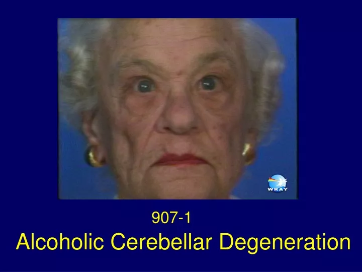
907-1. Alcoholic Cerebellar Degeneration. Clinical Syndrome. The clinical syndrome of alcoholic cerebellar degeneration is remarkably stereotyped. The usual presentation, as in this patient, is a progressive unsteadiness in walking evolving over months and years. Clinical Syndrome.

E N D
907-1 Alcoholic Cerebellar Degeneration
Clinical Syndrome The clinical syndrome of alcoholic cerebellar degeneration is remarkably stereotyped. The usual presentation, as in this patient, is a progressive unsteadiness in walking evolving over months and years.
Clinical Syndrome The cerebellar syndrome predominantly affects stance, eye movements, and gait, sometimes with trunkal ataxia and titubation. Dysarthria and upper limb ataxia are rare.
Pathophysiology Ataxia may develop during periods of abstinence. Identical cerebellar degeneration has been observed in non-alcoholic patients with severe malnutrition.
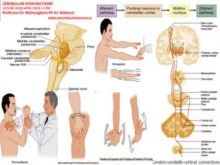
Eye Movements Square Wave Jerks Horizontal Saccadic Hypermetria Horizontal Gaze Evoked Nystagmus Saccadic Pursuit
Deficits Caused by Lesions of Dorsal Vermis, Fastigial Nucleus, and Uncinate Fasciculus Box 12-4. Leigh RJ, Zee DS. The Neurology of Eye Movements 4th Edition. Oxford University Press, New York 2006 with permission.
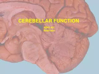
Pathological Changes Selective atrophy of the anterior and superior parts of the cerebellar vermis Involvement of the cerebellar hemispheres less extensive Loss of neurons in the cerebellum involves all types but Purkinje’s cells are the most seriously affected
MRI Figure 1: Sagittal T1WI shows striking atrophy of the superior vermis.
MRI Findings Figure 2: Axial T2WI through the upper midbrain and vermis shows the cerebellar folia are thinned and the CSF spaces increased. Courtesy Anne Osborn, M.D