Download
1 / 67
780 likes | 1.65k Views
Anemias in children. Anemia…. … abnormal low hemoglobin, hematocrit or RBC count, lower than the age-adjusted reference range for healthy children. Etiologic classification. I Impaired red cell formation A/ Deficiency Decreased dietary intake Increased demand

E N D
Anemia… … abnormal low hemoglobin, hematocrit or RBC count, lower than the age-adjusted reference range for healthy children.
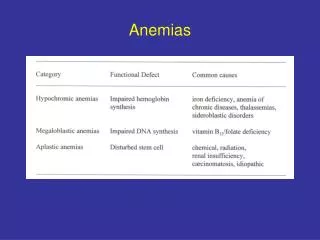
Etiologic classification I Impaired red cell formation A/ Deficiency • Decreased dietary intake • Increased demand • Decreased absorption • Increased loss B/ Bone marrow failure • Failure of a single or all cell lines • Infiltration C/Dyshematopoietic anemia II Blood loss III Hemolytic anemia • Corpuscular (membrane, enzymatic or hemoglobine defects) • Extracorpuscular (immune, idiopathic)
detailed history careful physical examination peripheral blood smear red cell morphology MCV RDW (red cell distribution width) WBC and platelet morphology Additionally: -bone marrow evaluation -additional testing Diagnosis of Anemia
History - Diet (iron , folate, vitB12 intake, onset of hemolysis after certain foods –e.g.,fava beans) - family history (transfusion requirements of relatives, splenectomy, gallblader disease) - environmental exposures (lead poissoning) - symptoms (headache, exertion dyspnea, fatigue,dizziness, weakness, mood or sleep disturbances, tinnitis) • melena, hematemesis, abdominal pain- chronic blood loss
Pallor(skin, oral mucosa, nail beds) Jaundice -hemolysis tachycardia tachypnea orthostatic hypotension venous hum systolic ejection murmur peripheral edema? Splenomegaly? Hepatomegaly? Glossitis? gingival pigmentation? Adenopathy? Facial, extremity examination Physical Examination
Peripheral Blood Componentsimportant! Different values dependent on age! • RBC • Hgb • HCT • MCV – 80 – 100 fl/L(a calculated value) • MCH • RDW • Reticulocyte Count
Low(<70 fl) *Hypochromic/Microcytic -Iron deficiency anemia -Thalassemia -Sideroblastic anemia -Chronic infection -Lead poisoning -Inborn errors of Fe metabolism -Severe malnutrition -Copper deficiency (>85fl) *Macrocytic Normal newborn Increased erythropoesis Post splenectomy Liver disease Aplastic anemia Megaloblastic anemia Down S. Obstructive jaundice MCV for Characterize Anemia
Normocytic Acute blood loss Infection Renal failure Connective tissue disorders Liver disease Disseminated malignancy Early iron deficiency Aplastic anemia Bone marrow infiltration Dyserythropoietic anemia
Causes -Dietary deficiency -Increased demand (growth) -Impaired absorption -Blood loss (menstrual problems) Symptoms GI: Anorexia, poor weight gain, pica, atrophic glossitis CNS: fatigue, irritability Cardiac: increased cardiac output, cardiac hypertrophy Dry skin, thin hair, pallor, nail ridges Iron Deficiency Anemia
*characteristics of peripheral blood smear microcytic hypochromic *MCV and Hgb– decreased (Hgb<12g/L) Ferritin – decreased (<13mg/dL) TIBC - high -Serum iron –decreased (N 50-150 µg/dL) Iron Deficiency Anemia
Iron Deficiency Anemia • Treatment • oral iron supplementation: 4 - 6mg/kg/day of elemental iron • goal: to replace iron stores, not just circulating Hgb! • Reticulocytes- starts to rise in 3 -4 days, • Hbg- after 4- 5 days • After Hgb normalisation – continue Fe therapy 1-2 months to replace Fe stores • *Iron- rich foods: animal protein, green vegetables, iron fortified cereales • Folate, vit C
parenteral therapy (IM,IV) • indications • poor compliance • severe bowel disease • intolerance of oral iron • chronic hemorrhage • acute diarrhea disorder
Megaloblastic anemia • Presence the megaloblasts in the bone marrow and macrocytes in the blood • In > 95% occurs as a result of folate and vitamin B12 deficiency • Deficiencies of ascorbic acid, tocopherol, thiamine may be related to megaloblastic anemia • Dietary vitamin B12 (cobalamine) is required from animal sources (meat and milk)
Causes of vitamin B12 deficiency IInadequate dietary intake (<2mg/day) –malnutrition, veganism, maternal deficiency II Defective vitamin B12 absorption • Failure to secrete intrinsic factor • Failure to absorption in small intestine III Defective vitamin B12 transport IV Disorders of vitamin B12 metabolism (congenital, acquired)
Folic acid deficiency • One of the most common micronutrient deficiences in the word (next to iron deficiency) • Component of malnutrition and starvation • Women are more frequently affected than men • Folate sufficiency prevents neural tube defects • Low mean daily folate intake is associated with twofold increased risk for preterm delivery and low infant birth weight
Causes of folic acid deficiency • Inadequate intake (method of cooking, special diet, goat’ milk) • Defective absorption (congenital or acquired) • Increased requirements (rapid growth, chronic hemolytic anemia, dyserythropoietic anemias, malignant disease, hypermetabolic state, cirrosis, post –BMT) • Disorders in folic acid metabolism (congenital, acquired) • Increased excretion
Clinical features of cobalamineand folate deficiency • Insidious onset: pallor, lethargy, fatigability, anorexia, sore red tongue and glossitis, diarrhea • History: similarly affected sibling, maternal vitamin B12 deficiency or poor maternal diet • Vitamin B12 deficiency: signs of neurodevelopmental delay, apathy, weakness, irrability, athetoid movements, hypotonia, peripheral neuropathy, spastic paresis
Diagnosis • Red cell changes: Hgb usually reduced, MCV increased to levels 110 – 140fl., MCHC normal, • in blood smear many macrocytes and macro-ovalocytes, anisocytosis, poikilocytosis, presence of Cabot rings, Howell-Jolly bodies, punctate basophilia • White blood cell count reduced to 1500 – 4000/mm3, neutrophils show hypersegmentation(>5 lobes) • Platelets count moderately reduced (50,000 – 180,000/mm3) • Bone marrow: megaloblastic appearance • Serum vitamin B12 values lowered (normal 200 – 800 pg/ml) • Serum and red cell folate levels – wide variation in normal range; less than 3 ng/ml -very low, 3-5 ng/ml –low, >5-6 ng/ml normal, in red cell:74-640 ng/ml • Schilling urinary excretion test – measurement of intrinsic factor availability and absorption of vitamin B12
Treatment Vitamin B12 deficiency Prevention in cases of risk of vitamin B12 deficiency Treatment 25 – 100µg vitamin B12 Folic acid deficiency Correction of the foliate deficiency (100-200µg/day) Treatment of the underlying causative disorder Improvement of the diet to increase folate intake
Isolated quantitative failure of one cell line, a single cytopenia , e.g. erythroid, myeloid, megakaryocytic • A failure of all three cell lines (pancytopenia with hypoplastic or aplastic bone marrow) • A quantitative failure of the bone marrow, e.g. congenital dyserythropoietic anemia • The invasion of the bone marrow by non-neoplastic or neoplastic condition
Diamond-Blackfan anemiacongenital pure red cell aplasia The erythroid progenitor cell is intrinsically abnormal in the following aspects: • Decreased sensitivity to erythropoietin (EPO) • Decreased sensitivity to EPO not corrected by IL-3 and GM-CSF caused by: • Functional abnormalities in the erythropoietin receptors • Erythroid progenitors are abnormally sensitive to a deprivation of erythropoietin, resulting in an accelerated rate of apoptosis
Clinical features • Anemia and pallor in first 3 months, 35 % is anemic at birth, 65% -identified by 6 months of age and 90% - by 1 year • Platelets and white cell count – normal • 25% have prenatal or postnatal growth failure and associated congenital defects, including short stature, abnormalities of thumbs, skeletal anormalities, congenital heart defects, webbed neck, urinary tract abnormalities and craniofacial dysmorphism • Chromosomal studies generally normal • No hepatosplenomegaly • Malignant potential (increased incidence of ALL, AML, hepatocellular carcinoma)
Diagnosis • Anemia and reticulocytopenia • Bone marrow with virtual absence of normoblasts Differential diagnosis • Transient erythroblastopenia of childhood (TEC) • Congenital hypoplastic anemia
Treatment • Prednisone 2 mg/kg/day, when the hemoglobin level reaches 10.0g/dl → dose reduction to minimum necessary • Packed red cell transfusion, leukocyte –depleted • Bone marrow transplantation in steroid –resistant, transfusion-dependent patients
Fanconi anemiacongenital aplastic anemia • Rare inherited disorder, autosomal-recessive trait • Pancytopenia: develops between 4 and 12 years of age • It may present with isolated anemia or leukopenia or anemia + thrombocytopenia • Macrocytosis (high MCV), high HbF, high erythropoetin, presence of i antigen – characteristic of stress erythropoiesis • Diepoxybutane (DEB)-induced chromosomal breakages • Hypocellularity and fatty replacement in bone marrow • congenital anomalies: patchy brown pigmentation of the skin, short stature, skeletal anomalies, hyperreflexia, hypogenitalism, microcephaly, microphthalmia, strabismus, ptosis, nystagmus, abnormalities of the ears, deafness, mental retardation, renal and cardiac anomalies • Chromosomal breakages and structural abnormalities, chromatoid exchange • High incidence of AML, carcinoma
Treatment: Supportive: • Packed red blood cells and platelets (irradiated, leukocyte reduced) • Chelation treatment in iron overload • Androgen therapy Active: • Allogenic bone marrow transplantation
Acquired aplastic anemiapathophysiology • Immunologically mediated, tissue-specific, organ-destructive mechanism • Exposition to an antigen → cells and cytokines of the immune system destroy stem cells in the marrow → pancytopenia • Gamma –interferon plays a central role in the pathophysiology of AA • T cells from AA patients secrete gamma-IFN and TNF – potent inhibitors of both early and late hematopoietic progenitor cells • Cytotoxic T cells secrete also IL-2, which causes polyclonal expansion of the T cells
Causes of acquired AA Idiopathic (70%) Secondary: • Drugs: cytostatics, antibiotics (sulfonamides, chloramphenicol), anticonvulsants (hydantoin), antirheumatics, antidiabetics, antimalarian • Chemicals: insecticides • Toxins: benzene, carbon tetrachloride, glue, toluene • Irradiation • Infections: viral (hepatitis A, B, C, HIV, EBV, CMV, parvovirus) • Immunologic disorders: GvHD • Preleukemia, MDS, thymoma • Malnutrition • Paroxysmal nocturnal hemoglobinuria
Severity • Severe AA: bone marrow cellularity <25% Granulocyte count <500/mm3 platelet count <20,000/mm3 reticulocyte count <40,000/mm3 • Very severe AA: granulocyte count <200/mm3
Clinical findings: • Anemia (pallor, easy fatigability, loss of appetite) • Thrombocytopenia ( petechiae, easy bruising, severe nosebleeds) • Leukopenia (increased susceptibility to infections and oral ulcerations) • Hyperplastic gingivitis • No: hepatosplenomegaly and lymphadenopathy
Laboratory findings • Anemia normocytic, normochromic • Reticulocytopenia • Leukopenia: granulocytopenia often < 1500/mm3 • Thrombocytopenia: often < 30,000/mm3 • Bone marrow: marked depression or absence hematopoietic cells and replacement by fatty tissue • Normal chromosomal analysis
Treatment Severe AA: • Allogeneic BMT • In the absence of availability of an HLA-matched sibling marrow donor - immunoablation ( ATG, cyclosporine, methylprednisolone, growth factors- G-CSF)
Corpuscular defects →Membrane defects →Enzyme defects →Hemoglobin defects →Congenital dyserythropoietic anemias Extracorpuscular defects →Immune →Nonimmune
Clinical features suggesting a hemolytic process • Ethnic factors: incidence of sickle gene factor in the black population (8%), high incidence of thalassemia in people of Mediterranean ancestry, high incidence of glucose-6-phosphate dehydrogenase deficiency among Sephardic Jews • Age factors: anemia and jaundice in an Rh+ infant born to a mother Rh- or a group A or group B infant born to a group 0 mother • History of anemia, jaundice, or gallstones in family • Persistent or recurrent anemia associated with reticulocytosis • Anemia unresponsive to hematinics • Intermittent bouts or persistent indirect hyperbilirubinemia • Splenomegaly • Hemoglobinuria • Presence of multiple gallstones
Corpuscular hemolytic anemias Membrane defects • Morphologic abnormalities: hereditary spherocytosis,elliptocytosis, stomatocytosis, acanthocytosis • Spectrin is responsible for maintaining red cell shape, regulates the lateral mobility of integral membrane proteins and provides structural support for the lipid bilayer
Hereditary spherocytosis Genetics • Autosomal-dominant inheritance (75%), non-family history –25% • Most common in people of northern European heritage • Incidence of 1 in 5000 Pathogenesis • Membrane instability due to dysfunction or deficiency of a red cell skeletal protein: ankyrin (75-90%) and/or spectrin (50%)
The sequelae are as follow: • Sequestration of red cells in the spleen (due to erythrocyte deformability) • Depletion of membrane lipid • Decrease of membrane surface area relative to volume, resulting in a decrease in surface area-to-volume ratio • Tendency to spherocytosis • Influx and efflux of sodium increased; cell dehydratation • Increased glycolysis • Premature red cell destruction
Hematology • Anemia mild to moderate; in erythroblastopenic crisis Hb may drop to 2 – 3g/dl • MCV usually decreased, MCHC raised • Reticulocytosis • Blood film – microspherocytes, hyperdense cells , polychromasia • Coomb’s test negative • Increased red cell osmotic fragility – spherocytes lyse in higher concentrations of saline than normal red cells, occasionally only demonstrated after incubation of blood sample at 37 C for 24 hours • Autohemolysis at 24 and 48 hours increased, corrected by the addition of glucose • Reduced red cell survival • Marrow- normoblastic hyperplasia, increased iron • EMA-test
Biochemistry • Raised bilirubin, mainly indirect reacting • Obstructive jaundice with increased direct-reacting bilirubin; may develop due to gallstones, a consequence of increased pigment excretion
Clinical features • Anemia and jaundice- severity depends on rate of hemolysis, degree of compensation of anemia by reticulocytosis, and ability of liver to conjugate and excrete indirect hyperbilirubinemia • Splenomegaly • Presents in newborn (50% of cases) with hyperbilirubinemia, reticulocytosis, normoblastosis, spherocytosis, negative Coomb’s test, and splenomegaly • Presence before puberty in most patients • Sometimes diagnosis made much later in life by chance
Complications • Hemolytic crisis – with pronounced jaundice due to accelerated hemolysis ( may be precipitated by infection) • Erythroblastopenic crisis – dramatic fall in Hb level and reticulocyte count, usually associated with parvovirus B19 infection • Folate deficiency caused by increased red cell turnover, may lead to superimposed megaloblastic anemia • Gallstones in 50% of untreated patients, incidence increases with age • Rarely hemochromatosis
Treatment • Folic acid supplement 1mg/day • Leukocyte-depleted packed red cell transfusion for severe erythroblastopenic crisis • Splenectomy for moderate to severe cases
Hereditary elliptocytosis (HE) • Is due to various defects in the skeletal proteins, spectrin and protein 4.1 , it results increased membrane rigidity and in decreased cellular deformability • Autosomal-dominant mode of inheritance • Elliptocytes varies from 50 to 90% • Osmotic fragility normal or increased • Treatment: transfusion, splenectomy, prophylactic folic acid
Another types of membrane defects • Hereditary stomatocytosis (the cells contain high Na and low K concentrations) • Hereditary acanthocytosis • Hereditary xerocytosis
Enzyme defects • Pyruvate Kinase deficiency:defective red cell glycolysis • Red cell rigid, deformed and metabolically and physically vulnerable • Autosomal –recessive inheritance • Nonspherocytic hemolytic anemia • Variable severity: moderate severe anemia • Neonatal jaundice • Splenomegaly • Gallstones, hemosiderosis, bone changes Treatment: folic acid supplementation, transfusions, splenectomy
Glucose-6-Phosphate Dehydrogenase deficiency • Sex-linked recessive mode of inheritance • Disease fully expressed in hemizygous males and homozygous females • Most frequent among blacks and those of Mediterranean origin • Associations : hemolysis may be produced by drugs, fava (broad) bean, infections
Clinical features Drug induced hemolysis : -Analgetics and antipyretics -Antimalarian agents -Sulfonamides -Nitrofurans -Sulfones Favism: -acute life-threating hemolysis often leading to acute renal failure caused by ingestion of fava beans Associated with mediterranean and Canton varieties Neonatal jaundice Chronic nonspherocytic anemia Treatment: Avoid drugs deleterious in G6PD, splenectomy