Download
1 / 30
330 likes | 594 Views
16. Innate Immunity : Nonspecific Defenses of the Host. SLOs. Differentiate between innate and adaptive immunity. Define toll-like receptors. Differentiate physical from chemical factors, and list examples of each. Describe the role of normal microbiota in innate resistance.

E N D
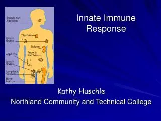
16 Innate Immunity: Nonspecific Defenses of the Host
SLOs Differentiate between innate and adaptive immunity. Define toll-like receptors. Differentiate physical from chemical factors, and list examples of each. Describe the role of normal microbiota in innate resistance. Classify phagocytic cells, and describe the roles of granulocytes and monocytes. Define and explain phagocyte and phagocytosis. Explain the different stages of inflammation. Describe the cause and effects of fever. Describe two of the three pathways of activating complement and describe the 3 outcomes. Compare and contrast the actions of -IFN and -IFN with -IFN. Describe the role of transferrins and antimicrobial peptides in innate immunity.
Cytokines! TLRs on Ms, dendritic cells, epithelial cells PAMPs recognition
Horseshoe structure of TLR3, showing attached sugars (spheres) and internal structures Fig. 16.7
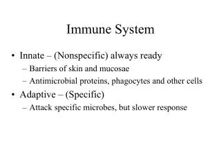
The Concept of Immunity • Susceptibility: Lack of resistance to a disease. • Immunity: Ability to ward off disease. • Innate immunity: Defenses against any pathogen. • Adaptive immunity: Immunity, resistance to a specific pathogen. Fig 16.1
First Line of Defense: Skin and Mucous Membranes Physical Factors • Epidermis: consists of tightly packed cells with keratin, a protective protein • Two other protective physical factors of skin? • Mucus of mucous membranes • Lacrimal apparatus • Saliva • Nose hairs • (Muco)-ciliary escalator Fig 16.3
Chemical Factors • Fungistatic fatty acids in sebum • Low pH (3-5) of skin • Lysozyme in _______________________ • Low pH (?) of gastric juice • Transferrins in blood Also important: Antagonism and competitive exclusion of normal microbiota
1st Line Defense in Human ANIMATION Host Defenses: The Big Picture
Second Line of Defense: Formed Elements in Blood Compare to Table 16.1 60-70% 2-4% 0.5-1%% 3-8% 20-25%
Process of Phagocytosis Phagocytes engulf and kill microorganisms Steps of phagocytosis: • Chemotaxis • Recognition and attachment • Engulfment and creation of phagosome • Fusion of phagosome with lysosome • Destruction and digestion • Residual body Exocytosis Fig 16.7
Phagocytosis Foundation Fig 16.7
Phagocytosis and Evasion of Phagocytosis ANIMATION Phagocytosis: Overview ANIMATION Phagocytosis: Mechanism Review the Following Textbook Animations ANIMATION Virulence Factors: Hiding From Host Defenses ANIMATION Virulence Factors: Inactivating Host Defenses ANIMATION Phagocytosis: Microbes That Evade It
Inflammation Tissue damage leads to inflammatory response Purpose: • Destroy pathogen • limit spread of infection • pave way for tissue repair 4 cardinal signs:? Acute-phase proteins (Chemical mediators) activated: • Complement proteins • Cytokines • Specialized proteins such as fibrinogen and bradykinin
The Three Stages of Inflammation • Vasodilation and increased vessel permeability due to histamine (and other cytokine) release edema • Phagocyte migration and phagocytosis • Margination and diapedesis (emigration) • Chemotaxis(due to various cytokines and components of complement system) • Pus formation • Factors challenging effectiveness of phagocytosis • Tissue repair and regeneration depends on type of tissue
Inflammatory Process Margination Diapedesis Compare to Fig 16.8
Fever: Abnormally High Body Temperature • Hypothalamus acts as body’s thermostat • Endotoxin causes phagocytes to release interleukin–1 (IL–1). IL-1 is an endogenous pyrogen • Hypothalamus releases prostaglandins that reset the thermostat • Body reacts to raise the temperature. How? • When no more IL–1, body temperature falls (crisis).
Beneficial effects of moderate fever: Inhibited pathogen growth Increased cellular metabolism e.g.: • Increased transferrin production • Increased IL–1 activity T cell production • Faster repair mechanisms Problematic effects of high fever: > 40.7C (105F) can be dangerous (Tachycardia, acidosis, dehydration) Death at temp. > 44 - 46C
Antimicrobial Substances • The complement system • Interferons • Transferrins: bind serum iron • Antimicrobial peptides: cause bacterial cell lysis. Produced by mucous membrane cells and phagocytes.
TheComplement System Compare to Foundation Fig 16.9
Complement System Summary Series of 30 plasma (serum) proteins, activated in a cascade Three effects of complement system: • Enhances inflammatory response, e.g.: attracts phagocytes • Increases phagocytosis through opsonization or immune adherence • Creates Membrane Attack Complexes (MACs) Cytolysis
Opsonins (complement proteins or antibodies) coat bacteria and promote attachment of micro-organism to phagocyte Opsonization
Classical Pathway Fig 16.12
Alternative Pathway Does not require a specific antibody to get started Fig 16.13
Some Bacteria Evade Complement • Capsules prevent Complement activation. • Surface lipid-carbohydrates of some Gram-negatives prevent MAC formation. • Enzymatic digestion of C5a by Gram-positives. ANIMATION Complement System: Overview ANIMATION Complement System: Activation ANIMATION Complement System: Results
Interferons (IFNs) • Family of glycoproteins • Host-cell-specific but not virus-specific • -IFN and -IFN: Produced by virus infected cells. Mode of action is to induce uninfected cells to produce antiviral proteins (AVPs) that inhibit viral replication. • -IFN: Produced by lymphocytes. Causes neutrophils and macrophages to phagocytize bacteria. Also involved in tumor immunology. • Recombinant interferons have been produced. However short-acting and many side-effects. No effect on already infected cells.
Interferons (IFNs) Fig 16.15
Applications of Microbiology: Serum Collection Unnumbered Figure 16.1a
Unnumbered Figure 16.1b the end