Download
1 / 24
260 likes | 353 Views
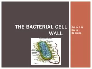
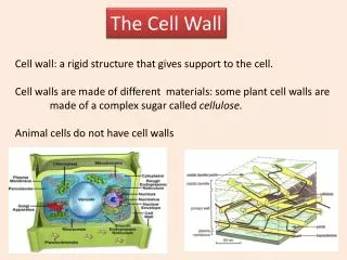
Structure of the Cell Wall. Helps determine the shape of a bacterium Provides strong structural support Most are rigid because of peptidoglycan content. Figure 4.13. Structure of the Cell Wall, cont. Keeps cells from rupturing because of changes in pressure due to osmosis

E N D
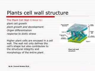
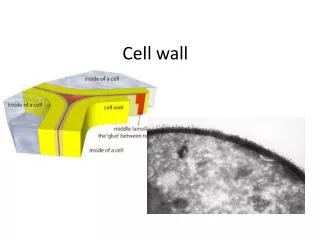
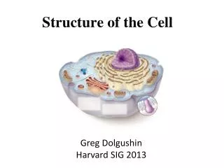
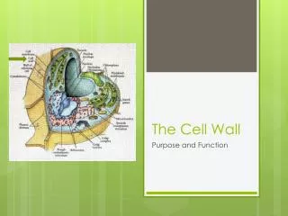
Structure of the Cell Wall • Helps determine the shape of a bacterium • Provides strong structural support • Most are rigid because of peptidoglycan content
Structure of the Cell Wall, cont. • Keeps cells from rupturing because of changes in pressure due to osmosis • Target of many antibiotics- disrupt the cell wall, and cells have little protection from lysis • Gram-positive cell wall • A thick (20 to 80 nm), homogeneous sheath of petidoglycan • Contains tightly bound acidic polysaccharides • Gram-Negative Cell Wall • Single, thin (1 to 3 nm) sheet of peptidoglycan • Periplasmic space surrounds the peptidoglycan
Nontypical Cell Walls • Some aren’t characterized as either gram-positive or gram-negative • Some don’t have a cell wall at all • For example, Mycobacterium and Nocardia- unique types of lipids • Archae- unusual and chemically distinct cell walls • Mycoplasmas- lack cell wall entirely
Mycoplasmas and Other Cell-Wall-Deficient Bacteria • Mycoplasma cell membrane is stabilized by sterols and is resistant to lysis • Very small bacteria (0.1 to 0.5 µm) • Range in shape from filamentous to coccus • Not obligate parasites • Can be grown on artificial media • Found in many habitats • Important medical species: Mycoplasma pneumonia
Some bacteria lose their cell wall during part of their life cycle • L-forms • Arise naturally from a mutation in the wall-forming genes • Can be induced artificially by treatment with a chemical that disrupts the cell wall • When this occurs with gram-positive cells, the cell becomes a protoplast • With gram-negative cells, the cell becomes a spheroplast
The Gram-Negative Outer Membrane • Similar to the cell membrane, except it contains specialized polysaccharides and proteins • Uppermost layer- contains lipopolysaccharide • Innermost layer- phospholipid layer anchored by lipoproteins to the peptidoglycan layer below • Outer membrane serves as a partial chemical sieve • Only relatively small molecules can penetrat • Access provided by special membrane channels formed by porin proteins
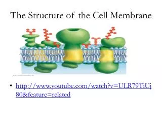
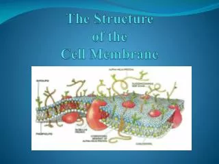
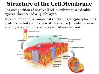
Cell Membrane Structure • Also known as the cytoplasmic membrane • Very thin (5-10 nm) • Contain primarily phospholipids and proteins • The exceptions: mycoplasmas and archae • Functions • Provides a site for functions such as energy reactions, nutrient processing, and synthesis • Regulates transport (selectively permeable membrane) • Secretion
Practical Considerations of Differences in Cell Envelope Structure • Outer membrane- an extra barrier in gram-negative bacteria • Makes them impervious to some antrimicrobial chemicals • Generally more difficult to inhibit or kill than gram-positive bacteria • Cell envelope can interact with human tissues and cause disease • Corynebacterium diphtheriae • Streptococcus pyogenes
Bacterial Internal Structure • Contents of the Cell Cytoplasm • Gelatinous solution • Site for many biochemical and synthetic activities • 70%-80% water • Also contains larger, discrete cell masses (chromatin body, ribosomes, granules, and actin strands)
Bacterial Chromosome • Single circular strand of DNA • Aggregated in a dense area of the cell- the nucleoid Figure 4.17
Plasmids • Nonessential pieces of DNA • Double-stranded circles of DNA • Often confer protective traits such as drug resistance or the production of toxins and enzymes
Ribosomes • Made of RNA and protein • Special type of RNA- ribosomal RNA (rRNA) • Characterized by S units- the prokaryotic ribosome is 70S Figure 4.18
Inclusions • Inclusions- also known as inclusion bodies • Some bacteria lay down nutrients in these inclusions during periods of nutrient abundance • Serve as a storehouse when nutrients become depleted • Some enclose condensed, energy-rich organic substances • Some aquatic bacterial inclusions include gas vesicles to provide buoyancy and flotation
Granules • A type of inclusion body • Contain crystals of inorganic compounds • Are not enclosed by membranes • Example- sulfur granules of photosynthetic bacteria • Polyphosphate granules of Corynebacterium and Mycobacterium are called metachromatic granules because they stain a contrasting color in methylene blue • Magnetotactic bacteria contain granules with iron oxide- give magnetic properties to the cell
The Actin Cytoskeleton • Long polymers of actin • Arranged in helical ribbons around the cell just under the cell membrane • Contribute to cell shape
Bacterial Endospores: An Extremely Resistant Stage • Dormant bodies produced by Bacillus, Clostridium, and Sporosarcina Figure 4.21
Endospore-Forming Bacteria • These bacteria have a two-phase life cycle • Phase One- Vegetative cell • Metabolically active and growing • Can be induced by the environment to undergo spore formation (sporulation)
Phase Two: Endospore • Stimulus for sporulation- the depletion of nutrients • Vegetative cell undergoes a conversion to a sporangium • Sporangium transforms in to an endospore • Hardiest of all life forms • Withstand extremes in heat, drying, freezing, radiation, and chemicals • Heat resistance- high content of calcium and dipicolinic acid • Some viable endospores have been found that were more than 250 million years old