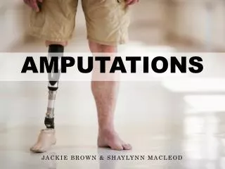
Download
1 / 19
310 likes | 905 Views
Forefoot Amputations. Dan Preece DPM, R1 July 22, 2009. Statistics Foot ulceration with infection is one of the leading causes of hospitalization for patients with diabetes mellitus. A pproximately 15% of all patients with diabetes will develop a foot or leg ulceration at some point.

E N D
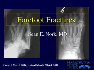
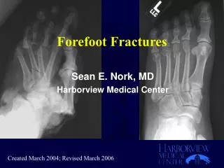
Forefoot Amputations Dan Preece DPM, R1 July 22, 2009
Statistics • Foot ulceration with infection is one of the leading causes of hospitalization for patients with diabetes mellitus. • Approximately 15% of all patients with diabetes will develop a foot or leg ulceration at some point. • Fifty percent of these ulcerations will recur within 18 months. • Diabetic patients are 15 times more likely to undergo a major limb amputation than patients without diabetes. • Approximately 50,000 amputations are performed yearly in patients with diabetes • Banks AS, Downey MS, Martin DE, Miller SJ, eds. McGlamry's Comprehensive Textbook of Foot and Ankle Surgery. 3rd edition. Philadelphia, PA: Lippincott, Williams &Wilkins; 2001. Pg 1567-1637.
Risk factors for Ulceration, Infection and Amputation: • Peripheral neuropathy • Structural foot abnormality • Limited joint mobility • Elevated, prolonged pressures • History of prior ulcers or amputation • Blindness or visual impairment • Chronic renal disease • Poor glycemic control • Duration of diabetes • Advanced Age Banks AS, Downey MS, Martin DE, Miller SJ, eds. McGlamry's Comprehensive Textbook of Foot and Ankle Surgery. 3rd edition. Philadelphia, PA: Lippincott, Williams &Wilkins; 2001. Pg 1567-1637.
Pecoraro et al. identified a triad of problems leading to 75% of the amputations in 80 pts. • Minor trauma such as accidental cuts, or shoe-related repetitive pressure that commonly results in the formation of corns and calluses. • Cutaneous ulceration arising from minor trauma. • Poor wound healing owing to the multiple complications associated with diabetes, such as peripheral vascular disease, neuropathy, hyperglycemia, and renal disease. • (Wound healing problems ultimately led to either infection or chronic wounds that required amputation for resolution.) Pecoraro RE, Reiber GE, Burgess EM. Pathways to limb amputation. Diabetes Care 1990;13:513-521.
Is It Osteomyelitis?? • Yuh et al. compared plain x-ray studies, bone scans, and magnetic resonance imaging (MRI) scans for specificity and sensitivity. He noted that they all have some limitations. • -MRI examination had the greatest sensitivity and specificity of these techniques, and is, by far, the most expensive. • Bone biopsy and culture remain the gold standard for diagnosis.
Probe to Bone = Osteo (or does it?) Grayson et al. (1995) Seventy-five patients with a total of 76 infected foot ulcers were studied. Osteomyelitis was diagnosed in 50 instances (66%) and was excluded in 26 instances. Palpating bone on probing the pedal ulcer had a sensitivity of 66% for osteomyelitis, a specificity of 85%, a positive predictive value of 89%. Results were skewed because all ulcers were clinically infected. Grayson ML, Gibbons GW, Balogh K, Levin E, Karchmer AW. Probing to bone in infected pedal ulcers. A clinical sign of underlying osteomyelitis in diabetic patients. JAMA 1995 Mar 1;273(9): 721-3. Jeffcoate, et. al. (2006) In a study of 104 outpatient foot ulcers, found a probe to bone test sensitivity of 38 percent and a specificity of 91 percent in correlation to the confirmed cases of osteomyelitis. All ulcers were included including non-infected. Shone A, Burnside J, Chipchase S, Game F, Jeffcoate W. Probing the validity of the probe-to-bone test in the diagnosis of osteomyelitis of the foot in diabetes.Diabetes Care. 2006 Apr;29(4):945.
Surgical Goals to Reduce Risk ofReulcerationand Amputation: • Reduce deformities such as: hammertoes, bunions, gastro-solealequinus, severe forefoot varus or valgus, forefoot equinus, charcot collapse etc… • Provide a stable/plantargrade foot for ambulation • Create a safely “shoe-able” foot.
Surgical Considerations: • Deep cultures from the bone • Bone biopsy • As much of the infected bone should be excised as possible • Soft, hemorrhagic bone should be resected as necessary to reach normal- appearing cortical or cancellous bone • Bonecan also be sent to pathology for microscopic evaluation to determine whether there has been a complete resection of the infected bone
Digital Amputations: -Control infection first, then amputate and close primarily? (May help increase amount of viable soft tissue for closure.) Or -Amputate immediately, control infection and close by delayed primary closure? (The answer may be a matter of preference or based on the stage of the infection. The more advanced the infection the more likely to amputate earlier.) • Take one toe or disarticulate/transmet head amputation? Often after removing a hallux, the lesser digits deviate and become plantarly prominent causing ulceration and infection. • If this is the second time around, a TMA should be considered.
Surgical approach: • - Incision should be planned only in viable tissue • - A converging semi-elliptic incision is made around the toe with anapex planned dorsally and plantarly. • Matchboth medial and lateral aspects of the incision to minimize remodeling of skin flaps following the initial incision. • Maximize one flap if an ulcer must be avoided on the other flap.
Neuropathy and Amputations: Often begins with the longest nerves. This may mean the hallux becomes insensate first, ulcerates, developesosteo and is amputated before the neuropathy is truly appreciated. The 2nd digit may follow suit and then the 3rd. Make sure your exam is thorough with the primary cause of ulceration being the top question in your mind. Source: Dr. Young
Single Ray Resection: • Amputation of a toe and its metatarsal is performed for: • localized abscess • osteomyelitis • necrotizing fasciitis extending along a single ray.
5th Ray Considerations: • -If amputation includes the base of the 5th met, consider the implications of losing the Per. Brevis and tertious… • -May lead to varus deformities, hallux limitus and bunions. • If the forefoot has a varus deformity and is not addressed, the 4th ray will be next to come off. Always verify that the 1st and 5th ray are bearing weight equally.
TMA: The incision should allow for a flap of thicker plantar skin to be brought dorsally. Bone cuts should mimic the parabola formed by the met heads. Shoe fillers should be created. Toe filler insert for TMA foot.
TMA Considerations: • -Most common site of new onset ulceration in a TMA pt will be styloidprocess, 5thmet head and the distal plantar lateral forefoot. Why? • The Achilles and Tibialis Anterior now invert the forefoot more so than without the opposing EDL . Plantar flexion is also increased. • The lateral mets are completely weightbearing from prox to distal, the more medial mets are only weight bearing at the heads, if you cut the heads off, the prox shaft may float in the air while the lateral mets bear even more weight. • TAL , STAT or Hibbs procedures should definitely be high on your list of surgical options to help control equino/varus forces.
Alternative to the Bone Saw: • Seidel C, et al. Drug therapy of diabetic neuropathic foot ulcers: transvenous retrograde perfusion versus systemic regimen. Vasa. 1991;20(4):388-93. • An isotonic saline solution containing gentamycin, buflomedil, dexamethasone, heparin and lignocain is injected into a dorsal foot vein under arterial occlusion of the lower leg. In the present study RVP treatment (1/90-12/90) was done in 20 patients with resistant DM ulcerations. Results were compared to a control group (CG) treated with systemic i.v. infusions (n = 20). • 10 days of treatment ulcers were closed in 6 vs. 0 (CG) patients, size diminuted in 10 vs. 3. • Non responders were not observed under RVP in contrast to Control G (7/20 cases). • In 4 of 5 RVP patients with secondary osteomyelitis, some restoration of osteolytic lesions was seen and none of 7 CG cases. • Rate of toe amputation dropped from 20% (CG) to 0%, • mean time of hospitalization was cut by 7 days in the RVP group. • Planimetry of the ulcered areas confirmed remarkable diminution in the RVP group. • Considering the striking differences between either regimen, RVP can be recommended for treatment of DNPU especially when complicated by osteomyelitis.
Alternative to the Bone Saw: • Agarwal P, Agrawal PK, Sharma D, Baghel KD J. Intravenous infusion for the treatment of diabetic and ischaemic non-healing pedal ulcers. EurAcad Dermatol Venereol. 2005 Mar;19(2):158-62. • Assessed the role of retrograde venous perfusion (RVP) for the treatment of nine diabetic and 10 ischaemic non-healing pedal ulcers. • Agents used were soda bicarbonate, heparin, lignocaine, gentamicin and pentoxiphylline(trental). • Five of nine diabetic non-healing ulcers showed complete healing and the remaining four improved. • The complete recovery in the cases of diabetic ulcer occurred in 10-24 days (mean 16 days), while ischemic ulcers took 10-14 days for complete recovery (mean 13.6 days). • There was a reduction of rest pain in all 10 patients with ischaemic disease; five patients showed complete healing of ulcers, and the other five improved significantly. • In two patients, pre-gangrene changes were reversed. • RVP is a useful adjunct to conservative or surgical treatment of non-healing pedal ulcers. Its main impact was in improving ischaemia and promoting healing.
References: • Banks AS, Downey MS, Martin DE, Miller SJ, eds. McGlamry's Comprehensive Textbook of Foot and Ankle Surgery. 3rd edition. Philadelphia, PA: Lippincott, Williams &Wilkins; 2001. Pg 1567-1637. • Pecoraro RE, Reiber GE, Burgess EM. Pathways to limb amputation. Diabetes Care 1990;13:513-521. • Yuh WT, Corson JD, Baraniewski HM, Rezai K, et al. Osteomyelitis of the foot in diabetic patients: evaluation with plain film, 99mTc-MDP bone scintigraphy, and MR imaging.AJR Am J Roentgenol. 1989 Apr;152(4):795-800. • Grayson ML, Gibbons GW, Balogh K, Levin E, Karchmer AW. Probing to bone in infected pedal ulcers. A clinical sign of underlying osteomyelitis in diabetic patients. JAMA 1995 Mar 1;273(9): 721-3. • Shone A, Burnside J, Chipchase S, Game F, Jeffcoate W. Probing the validity of the probe-to-bone test in the diagnosis of osteomyelitis of the foot in diabetes.Diabetes Care. 2006 Apr;29(4):945. • Seidel C, et al. Drug therapy of diabetic neuropathic foot ulcers: transvenous retrograde perfusion versus systemic regimen. Vasa. 1991;20(4):388-93. • Agarwal P, Agrawal PK, Sharma D, Baghel KD J. Intravenous infusion for the treatment of diabetic and ischaemic non-healing pedal ulcers. EurAcad Dermatol Venereol. 2005 Mar;19(2):158-62.