Download
1 / 51
510 likes | 1.02k Views
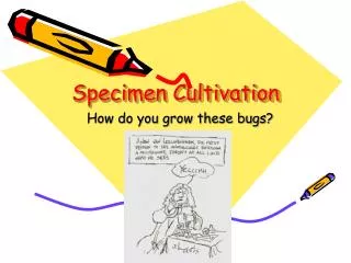
Specimen Cultivation. How do you grow these bugs?. Introductory remarks.

E N D
Specimen Cultivation How do you grow these bugs?
Introductory remarks • Although the future trends in clinical microbiology are clearly pointing in the direction of developing rapid, non growth dependent means of detecting the presence of infectious microorganisms (detection of microbial antigens, probing for genes, PCR), the isolation and identification of viable pathogens is still the major means by which infectious diseases are diagnosed today. This is particularly true for bacterial infections.
Introductory remarks • In order to identify an infectious bacterial agent by biochemical methods and to do antimicrobic sensitivity testing, it is necessary to have a pure culture of the organism. Why? • The way that we do this in the clinical lab is to streak a portion of the original sample out on primary isolation media to get isolated colonies.
Introductory remarks • Each isolated colony is a pure culture since it is theoretically the progeny of a single cell. • Each type of bacteria forms a characteristic colony in terms of shape, size, color, texture, and adherence to the medium. • These colonial characteristics are often used in the clinical lab as a first step in the process of identification of bacteria.
Introductory remarks • When the term culture media is used, it is important to keep in mind that it refers to artificial media on which bacteria and fungi are grown. • It is also important to remember that some bacteria have never been successfully grown on artificial lab media. For example: • Treponema pallidum – is usually grown in the testes of rabbits
Introductory remarks • Mycobacterium leprae – is usually grown in the nine-banded armadillo or in the foot-pads of mice. • Some bacteria (Rickettsia and Chlamydia) and all viruses are obligate intracellular parasites and can only be grown in living host cells, such as whole organisms or tissue culture cells. • For viruses grown in tissue culture, identification is often based on electron microscopic visualization of virus in the cells, or • Cytopathic effects (visible damage to or changes in the cells) produced by growth of the virus in the cells or by • Inclusion bodies • Syncytia formation • Rounding up of the cells
Introductory remarks • Growth of obligate intracellular parasites is very time consuming and expensive • Diagnosis of these organisms as the causative agent of an infection is usually by immunological means. • In a direct assay one looks for antigens from the organism. Therefore, one is looking for the organism, itself, usually in tissues. • In an indirect assay, one looks in the host for evidence of an immunological response against the organism, usually in the form of specific antibodies against the organism.
Introductory remarks • When trying to diagnose an infection by assaying for antibodies against the causative agent, it is best to take both an acute (when patient is the most ill) and a convalescent (when patient is recovering) phase specimen. Typically one looks for a 4-fold rise in antibody titer (amount of antibody/ml of sera) between acute and convalescent specimens. Why? • Parasites are not easily grown on artificial media or in living hosts. • The diagnosis of parasitic infections is usually based on the visual identification of the parasite itself or on microscopic identification of the egg, ova, or cyst produced by the parasite.
Plating of specimens and incubation of cultures for bacteria and fungi • When should the specimen be plated? • The specimen should be plated immediately because if there is a delay this may result in: • A loss of fastidious or anaerobic organisms or • There may be an overgrowth by normal flora in the specimen resulting in a change not only in the totalnumber of microorganisms, but also in the relative numbers of microorganisms. Why is this a problem?
Plating of specimens and incubation of cultures for bacteria and fungi • What should the specimen be plated on? • Diagnostic labs vary in their choices of routine plating media that is used for growing bacteria and fungi typically isolated from different types of specimens. • The choices used by different labs take into account what pathogens are anticipated in a given type of specimen, their growth requirements ( CO2, different temperature requirements, etc.) and the peculiarities of the available media.
Plating of specimens and incubation of cultures for bacteria and fungi • Types of media that may be used include: • General purpose media – this is media that supports the growth of most common pathogens and is therefore, classified as non-selective. However, in plate form, this type of media permits the isolation and differentiation of a wide variety of bacteria via differences in colony size, shape, color, texture, and adherence to the culture media. Examples of general purpose media include:
Plating of specimens and incubation of cultures for bacteria and fungi • Nutrient agar
Plating of specimens and incubation of cultures for bacteria and fungi • Blood agar –this media also allows differentiation based on the production by bacteria of hemolysins that destroy red blood cells in the agar. • Alpha () hemolysis is incomplete hemolysis and appears as a green halo surrounding the bacterial colony • Beta () hemolysis is complete hemolysis and appears as a clear area surrounding the bacterial colony • Gamma () hemolysis is no hemolysis at all
Types of hemolysis Alpha Beta Gamma
Plating of specimens and incubation of cultures for bacteria and fungi • Chocolate agar – is essentially the same as blood agar, except that the RBCs are lysed when added to the agar. This releases hemin and NAD for utilization by fastidious bacteria. The lysis gives the medium its chocolate brown color.
Plating of specimens and incubation of cultures for bacteria and fungi • Some books call blood agar and chocolate agar “enriched media” because the blood that is added to the media acts to “enrich” the media. • Selective media – this type of media contains special nutrients that support the growth of certain pathogens and/or inhibitors that suppress the growth of competing normal flora. For example: • Phenyl ethyl alcohol agar (PEA) – contains phenyl ethyl alcohol to inhibit the growth of Gram negative bacteria (Why does it inhibit the growth of G – bacteria?)
Plating of specimens and incubation of cultures for bacteria and fungi • Selective media is also usually differential. What makes it differential is often the addition of a carbohydrate and a pH indicator that together allow one to differentiate between organisms that ferment the carbohydrate and those that do not. For example: • Mannitol salts agar (MSA)- this media contains: • 7.5% NaCl to suppress the growth of organisms that are not halophilic (selection) • Mannitol and the pH indicator phenol (for differentiation). • If an organism that grows on the media ferments mannitol, acid is produced and this lowers the pH. At a low pH phenol red turns yellow. Therefore, organisms that ferment mannitol turn the media yellow:
Plating of specimens and incubation of cultures for bacteria and fungi • MacConkey agar (Mac) – this media contains: • Crystal violet to inhibit the growth of G+ bacteria and fungi (selection). • Lactose and the pH indicator neutral red which is red or pink at an acid pH (differentiation). • Organisms that are able to grow on the media and that ferment lactose produce pink colonies on the media • Organisms that don’t ferment lactose produce colorless colonies. • Mac plates are an example of what are collectively called enteric agar plates because they facilitate the isolation and differentiation of enteric pathogens. You will be discussing other types of enteric agar plates in the laboratory.
Plating of specimens and incubation of cultures for bacteria and fungi • Reducing media – this type of media is used for cultivating anaerobes. It contains compounds that chemically combine with dissolved oxygen in the media to deplete the O2 from the media. For example: • Sodium thioglycollate broth contains: • Thioglycolic acid as a reducing agent to create an anaerobic atmosphere deeper in the tube and • Resazurin as an oxygen-reduction indicator. In the presence of O2, resazurin turns pink. Where do different type of organisms grow in the thioglycollate broth?
Growth of different types of organisms in thioglycollate broth
Plating of specimens and incubation of cultures for bacteria and fungi • Enrichment culture media – is used to prevent missing a type of bacteria present in only small numbers. • This media is usually liquid and it provides nutrients and environmental conditions that favor the growth of one type of organism while being unsuitable for the growth of others. For example, to enrich a stool culture for enteric pathogens that may be found in low numbers relative to the normal flora from the intestine: • Gram negative broth • Tetrathionate broth • Selenite broth
Plating of specimens and incubation of cultures for bacteria and fungi • Special plating procedures • Anaerobic cultures • Reducing media may be used, or • Plates can be incubated in a special jar or pouch in an oxygen free atmosphere. In an anaerobic jar the oxygen free atmosphere is generated by the following mechanism:
Plating of specimens and incubation of cultures for bacteria and fungi • Envelopes containing sodium bicarbonate and borohydride are placed in the jar and water is added and the chemical reaction that occurs generates CO2 and H2. • The H2 that is generated combines with O2 in the jar in the presence of a palladium coated alumina pellet that acts as a catalyst for the following reaction: 2H2 + O22H2O Thus O2 is removed from the atmosphere. • An indicator strip with methylene blue that is colorless in the absence of oxygen and blue in the presence of oxygen is also placed in the jar.
Plating of specimens and incubation of cultures for bacteria and fungi • Blood cultures – as discussed previously, blood cultures are usually inoculated into blood culture media directly at the bedside of the patient. • Frequently two bottles of liquid media are inoculated, one for aerobic growth (TSB – tryptic soy broth) and one for anaerobic growth (Thio). • The bottles are usually examined every day for turbidity for 7-14 days. • If turbidity develops, some of the media is removed for Gram staining and subculturing onto solid media. • Depending upon the lab policies, blood cultures may routinely be Gram stained and subcultured at specific intervals (24 hrs.,48 hrs, etc.) even in the absence of turbidity.
Plating of specimens and incubation of cultures for bacteria and fungi • Some labs have machines (Bactec) that automatically detect growth in blood cultures by CO2 production • Quantitative counts – as discussed previously, this is often done on urine specimens. • A known volume of specimen is plated on the agar medium, via the use of a calibrated loop, and the number of colonies that grow are counted. • Caution – this only represents the number of bacteria present at the moment of plating. • For a clean catch urine specimen >100,000 colonies /ml is considered significant and indicative of disease. • For a bladder or kidney specimen >10,000 colonies/ml is considered significant and indicative of disease. Why?
Plating of specimens and incubation of cultures for bacteria and fungi • Plating for unusual bacteria – • Some of the more rarely encountered pathogens need unusual media and/or techniques to facilitate their isolation. • If a physician suspects that one of these organisms is the causative agent of an infection, he/she must notify the lab so that appropriate media can be prepared and proper precautions can be taken, if necessary. For example: • Brucella species (sp.) • Bordetella sp. • Legionella sp.
Plating of specimens and incubation of cultures for bacteria and fungi • Incubation of cultures • Temperature • Media inoculated with human specimens is usually incubated at 35-370 C, which is the optimum growth temperature for most human pathogens. • Fungi are often grown at room temperature (RT). • Many fungi are dimorphic and will grow as yeast at 370 C and as molds at RT. Candida albicans is different in that at RT it grows as a yeast, but at 370C andin the presence of serum, it grows as a mold.
Plating of specimens and incubation of cultures for bacteria and fungi • Atmosphere • Most pathogenic bacteria grow best at 2-10% CO2 and most clinical labs routinely use 5% CO2 incubators for their cultures. • Certain bacteria actually require 5-10% CO2 in order to grow or to grow well (Neisseria sp., Streptococcus sp., Haemophilus sp.). • To facilitate the growth of theses organisms, we will grow them in candle jars to provide the higher CO2 that they need for growth.
Plating of specimens and incubation of cultures for bacteria and fungi • Anaerobes have already been discussed • Length of incubation time • Most routine cultures are kept for 16-18 hours (overnight) before being reported out as negative • CSF and blood cultures are usually kept 1 week before being reported out as negative • Wound cultures may be kept for 48 hours before being reported out as negative