Download
1 / 51
540 likes | 731 Views
Chapter 6 Transcription. 王心宇 副教授 College of Life Sciences. 11.1 Introduction. Figure 11.1 The function of RNA polymerase is to copy one strand of duplex DNA into RNA. 11.1 Introduction.

E N D
Chapter 6 Transcription 王心宇 副教授 College of Life Sciences
11.1Introduction Figure 11.1The function of RNA polymerase is to copy one strand of duplex DNA into RNA.
11.1Introduction Figure 11.2A transcription unit is a sequence of DNA transcribed into a single RNA, starting at the promoter and ending at the terminator.
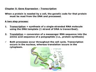
11.1Introduction • RNA polymerases are enzymes that synthesize RNA using a DNA template (formally described as DNA-dependent RNA polymerases). • A promoter is a region of DNA where RNA polymerase binds to initiate transcription. • Startpoint (startsite) (Startsite) refers to the position on DNA corresponding to the first base incorporated into RNA. • A terminator is a sequence of DNA that causes RNA polymerase to terminate transcription. • A transcription unit is the distance between sites of initiation and termination by RNA polymerase; may include more than one gene.
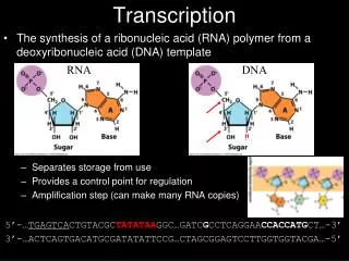
11.2Transcription occurs by base pairing in a "bubble" of unpaired DNA Figure 11.3DNA strands separate to form a transcription bubble. RNA is synthesized by complementary base pairing with one of the DNA strands.
11.2Transcription occurs by base pairing in a "bubble" of unpaired DNA Figure 11.4Transcription takes place in a bubble, in which RNA is synthesized by base pairing with one strand of DNA in the transiently unwound region. As the bubble progresses, the DNA duplex reforms behind it, displacing the RNA in the form of a single polynucleotide chain.
11.2Transcription occurs by base pairing in a "bubble" of unpaired DNA • RNA polymerase separates the two strands of DNA in a transient "bubble" and uses one strand as a template to direct synthesis of a complementary sequence of RNA. • The length of the bubble is ~12-14 bp, and the length of RNA-DNA hybrid within it is ~8-9 bp. Figure 11.5During transcription, the bubble is maintained within bacterial RNA polymerase, which unwinds and rewinds DNA, maintains the conditions of the partner and template DNA strands, and synthesizes RNA.
11.3The transcription reaction has three stages • RNA polymerase initiates transcription after binding to a promoter site on DNA. • During elongation the transcription bubble moves along DNA and the RNA chain is extended in the 5’ –3’ direction. • Transcription stops, the DNA duplex reforms and RNA polymerase dissociates at a terminator site. Figure 11.6Transcription has four stages, which involve different types of interaction between RNA polymerase and DNA. The enzyme binds to the promoter and melts DNA, remains stationary during initiation, moves along the template during elongation, and dissociates at termination.
11.3The transcription reaction has three stages Figure 11.7A stalled RNA polymerase can be released by cleaving the 3’end of the transcript.
11.4A model for enzyme movement is suggested by the crystal structure Figure 11.8The side view of the crystal structure of RNA polymerase II from yeast shows that DNA is held downstream by a pair of jaws and is clamped in position in the active site, which contains an Mg++ ion.
11.4A model for enzyme movement is suggested by the crystal structure Figure 11.9The end view of the crystal structure of RNA polymerase II from yeast shows that DNA is surrounded by ~270° of protein.
11.4A model for enzyme movement is suggested by the crystal structure Figure 11.11Movement of a nucleic acid polymerase requires breaking and remaking bonds to the nucleotides that occupy fixed positions relative to the enzyme structure. The nucleotides in these positions change each time the enzyme moves a base along the template.
11.4A model for enzyme movement is suggested by the crystal structure Figure 11.12The RNA polymerase elongation cycle starts with a straight bridge adjacent to the nucleotide entry site. After nucleotide addition, the enzyme moves one base pair and bridge bends as it retains contact with the newly added nucleotide. When the bridge is released, the cycle can start again.
11.5RNA polymerase consists of the core enzyme and sigma factor • The holoenzyme (complete enzyme) is the complex of five subunits including core enzyme (α2ββ’ ) and σ factor that is competent to initiate bacterial transcription. The β and β’ subunits together make up the catalytic center.The α subunit is required for assembly of the enzyme, and also plays a role in the interaction of RNA polymerase with some regulatory factors. • The core enzyme is the complex of RNA polymerase subunits that undertakes elongation. It does not include additional subunits or factors that may needed for initiation or termination. • Sigma factor is the subunit of bacterial RNA polymerase needed for initiation; it is the major influence on selection of promoters.
11.5RNA polymerase consists of the core enzyme and sigma factor The drug rifampicin (利福平) blocks transcription by bacterial RNA polymerase. It is a major drug used against tuberculosis(肺结核). Figure 11.13Both the template and coding strands of DNA are contacted by the β and β’ subunits largely in the region of the transcription bubble and downstream. The RNA is contacted mostly in the transcription bubble.
11.5RNA polymerase consists of the core enzyme and sigma factor • A loose binding site is any random sequence of DNA that is bound by the core RNA polymerase when it is not engaged in transcription. Figure 11.14Core enzyme binds indiscriminately to any DNA. Sigma factor reduces the affinity for sequence-independent binding, and confers specificity for promoters.
11.5RNA polymerase consists of the core enzyme and sigma factor • Sigma factor changes the DNA-binding properties of RNA polymerase so that its affinity for general DNA is reduced and its affinity for promoters is increased. • Binding constants of RNA polymerase for different promoters vary over 6 orders of magnitude, corresponding to the frequency with which transcription is initiated at each promoter.
11.6How does RNA polymerase find promoter sequences? How is RNA polymerase distributed in the cell? The main feature is that virtually all of it is bound to DNA, with no free core enzyme or holoenzyme. Figure 11.15Core enzyme and holoenzyme are distributed on DNA, and very little RNA polymerase is free.
11.6How does RNA polymerase find promoter sequences? Figure 11.16RNA polymerase binds very rapidly to random DNA sequences and could find a promoter by direct displacement of the bound DNA sequence.
11.6How does RNA polymerase find promoter sequences? • The rate at which RNA polymerase binds to promoters is too fast to be accounted for by random diffusion. • RNA polymerase probably binds to random sites on DNA and exchanges them with other sequences very rapidly until a promoter is found.
11.7Sigma factor controls binding to DNA Figure 11.17RNA polymerase passes through several steps prior to elongation. A closed binary complex is converted to an open form and then into a ternary complex.
11.7Sigma factor controls binding to DNA Figure 11.18Sigma factor and core enzyme recycle at different points in transcription.
11.7Sigma factor controls binding to DNA • When RNA polymerase binds to a promoter, it separates the DNA strands to form a transcription bubble and incorporates up to 9 nucleotides into RNA. • There may be a cycle of abortive initiations before the enzyme moves to the next phase. • Sigma factor may be released from RNA polymerase when the nascent RNA chain reaches 8-9 bases in length. • The change in association between sigma and holoenzyme changes binding affinity for DNA so that core enzyme can move along DNA.
11.8Promoter recognition depends on consensus sequences • A consensus sequence is an idealized sequence in which each position represents the base most often found when many actual sequences are compared. There are four (perhaps five) conserved features in a bacterial promoter: startpoint: purine, CAT. –10 sequence: The consensus is TATAAT. –35 sequence: The consensus is TTGACA separation between the –10 and –35 sequences: 16-18 bp in 90% of promoters
11.8Promoter recognition depends on consensus sequences Figure 11.19A typical promoter has three components, consisting of consensus sequences at -35 and -10, and the startpoint.
11.8Promoter recognition depends on consensus sequences Figure 11.20One face of the promoter contains most of the contact points for RNA. The initial region of unwinding extends from within the -10 sequence to past the startpoint.
11.8Promoter recognition depends on consensus sequences • A promoter is defined by the presence of short consensus sequences at specific locations. • The bacterial promoter consensus sequences consist of a purine at the startpoint, the hexamer TATAAT centered at –10, and another hexamer centered at –35. • Individual promoters usually differ from the consensus at one or more positions. • The consensus sequences at –35 and –10 provide most of the contact points for RNA polymerase in the promoter. • The points of contact lie on one face of the DNA.
11.9Promoter efficiencies can be increased or decreased by mutation • A down mutation in a promoter decreases the rate of transcription. • An up mutation in a promoter increases the rate of transcription.
11.9Promoter efficiencies can be increased or decreased by mutation • Down mutations to decrease promoter efficiency usually decrease conformance to the consensus sequences, whereas up mutations have the opposite effect. • Mutations in the –35 sequence usually affect initial binding of RNA polymerase. • Mutations in the –10 sequence usually affect the melting reaction that converts a closed to an open complex.
11.10Supercoiling is an important feature of transcription Transcription has a significant effect on the (local) structure of DNA. As a result, two enzymes, gyrase (which introduces negative supercoils) and topoisomerase I (which removes negative supercoils) are required to rectify the situation in front of and behind the polymerase, respectively. Figure 11.22Transcription may generate more tightly wound (positively supercoiled) DNA ahead of RNA polymerase, while the DNA behind becomes less tightly wound (negatively supercoiled).
11.11Substitution of sigma factors may control initiation Figure 11.23The sigma factor associated with core enzyme determines the set of promoters where transcription is initiated.
11.11Substitution of sigma factors may control initiation Figure 11.24In addition toσ70, E. coli has several sigma factors that are induced by particular environmental conditions. (A number in the name of a factor indicates its mass.)
11.11Substitution of sigma factors may control initiation Figure 11.25Transcription of phage SPO1 genes is controlled by two successive substitutions of the sigma factor that change the initiation specificity.
11.11Substitution of sigma factors may control initiation Figure 11.26E. coli sigma factors recognize promoters with different consensus sequences.
11.11Substitution of sigma factors may control initiation • E. coli has several sigma factors, each of which causes RNA polymerase to initiate at a set of promoters defined by specific –35 and –10 sequences. • σ70 is used for general transcription, and the other sigma factors are activated by special conditions. • A cascade of sigma factors is created when one sigma factor is required to transcribe the gene coding for the next sigma factor. • Sigma factor cascades are used to control transcription in some bacteriophage infections.
11.12Sigma factors directly contact DNA • σ 70 changes its structure to release its DNA-binding regions when it associates with core enzyme. Figure 11.27Sigma factor has an elongated structure that extends along the surface of the core subunits when the holoenzyme is formed.
11.12Sigma factors directly contact DNA Figure 11.28DNA initially contacts sigma factor (pink) and core enzyme (gray). It moves deeper into the core enzyme to make contacts at the –10 sequence. When sigma is released, the width of the passage containing DNA increases.
11.12Sigma factors directly contact DNA The use of α-helical motifs in proteins to recognize duplex DNA sequences is common. Amino acids separated by 3-4 positions lie on the same face of an α-helix and are therefore in a position to contact adjacent base pairs. Figure 11.29Amino acids in the 2.4 α-helix of σ70 contact specific bases in the coding strand of the –10 promoter sequence.
11.12Sigma factors directly contact DNA Figure 11.30The N-terminus of sigma blocks the DNA-binding regions from binding to DNA. When an open complex forms, the N-terminus swings 20 Å away, and the two DNA-binding regions separate by 15 Å.
11.13There are two types of terminators in E. coli There are two types of terminators in E. coli, and they are distinguished according to whether RNA polymerase requires any additional factors to terminate in vitro: • Core enzyme can terminate at certain sites in the absence of any other factor. These sites are called intrinsic terminators. • Rho-dependent terminators are defined by the need for addition of rho factor (ρ). ..
11.13There are two types of terminators in E. coli Termination may require both recognition of the terminator sequence in DNA and the formation of a hairpin structure in the RNA product. Figure 11.31The DNA sequences required for termination are located prior to the terminator sequence. Formation of a hairpin in the RNA may be necessary.
11.13There are two types of terminators in E. coli • Terminators vary widely in their efficiencies of termination. At some terminators, the termination event can be prevented by specific ancillary factors that interact with RNA polymerase. Antitermination causes the enzyme to continue transcription past the terminator sequence, an event called readthrough (the same term used to describe a ribosome's suppression of termination codons).
11.14Intrinsic termination requires a hairpin and U-rich region The role of the hairpin in RNA is probably to cause RNA polymerase to slow, thus creating an opportunity for termination to occur. A downstream U-rich region destabilizes the RNA-DNA hybrid when RNA polymerase pauses at the terminator hairpin. Figure 11.32Intrinsic terminators include palindromic regions that form hairpins varying in length from 7-20 bp. The stem-loop structure includes a G-C-rich region and is followed by a run of U residues.
11.15How does rho factor work? Rho-dependent terminators account for about half of E. coli terminators. The sequences required for rho-dependent termination are 50-90 bases long and lie upstream of the termination site. Their common feature is that the RNA is rich in C residues and poor in G residues. Figure 11.33 A rut site has a sequence rich in C and poor in G preceding the actual site(s) of termination. The sequence correspodns to the 3’end of the RNA.
11.15How does rho factor work? Rho factor is an essential protein in E. coli. It functions solely at the stage of termination. The subunit has an RNA-binding domain and an ATP hydrolysis domain. Figure 11.34Rho has an N-terminal RNA-binding domain and a C-terminal ATPase domain. A hexamer in the form of a gapped ring binds RNA along the exterior of the N-terminal domains. The 5’ end of the RNA is bound by a secondary binding site in the interior of the hexamer.
Figure 11.35Rho factor binds to RNA at a rut site and translocates along RNA until it reaches the RNA-DNA hybrid in RNA polymerase, where it releases the RNA from the DNA.
11.16Antitermination is a regulatory event Antiterminationis a mechanism of transcriptional control in which termination is prevented at a specific terminator site, allowing RNA polymerase to read into the genes beyond it. Figure 11.36Antitermination can be used to control transcription by determining whether RNA polymerase terminates or reads through a particular terminator into the following region.
11.16Antitermination is a regulatory event Antitermination proteins allow RNA polymerase to transcribe through certain terminator sites. Figure 11.37An antitermination protein can act on RNA polymerase to enable it to readthrough a specific terminator.
11.16Antitermination is a regulatory event antitermination event is not determined by the terminators (tL1 and tR1); the recognition site needed for antitermination lies upstream in the transcription unit, that is, at a different place from the terminator site at which the action eventually is accomplished. Figure 11.38Host RNA polymerase transcribes lambda genes and terminates at t sites. pN allows it to read through terminators in the L and R1 units; pQ allows it to read through the R’ terminator. The sites at which pN acts (nut) and at which pQ acts (qut) are located at different relative positions in the transcription units.
11.16Antitermination is a regulatory event Figure 11.39Ancillary factors bind to RNA polymerase as it passes the nut site. They prevent rho from causing termination when the polymerase reaches the terminator.