Download
1 / 47
470 likes | 573 Views
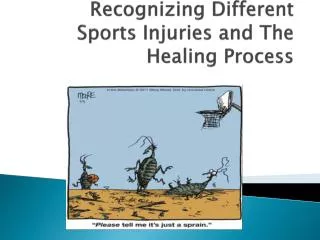
The Healing Process. Guidelines for Choosing the Proper Modality. Choosing the Proper Modality. How do you know what to use, and how do you know when to use it? Theoretical knowledge Practical experience

E N D
The Healing Process Guidelines for Choosing the Proper Modality
Choosing the Proper Modality • How do you know what to use, and how do you know when to use it? • Theoretical knowledge • Practical experience *You can’t follow the same recipe for every patient; avoid “cookie cutter” treatment plans
Modalities in a POC • Modalities should be an adjunct to TEs • ROM and strengthening TEs are the desired end goal • Rehab protocols and progressions must be based primarily on the physiologic responses of the tissues to injury and on an understanding of how various tissues heal • What does this mean? • The therapist must understand the healing process in order to choose the proper modality at the proper time
Modalities in a POC • Decisions on how and when modalities may be best used relies upon: • Recognition of signs and symptoms • Awareness of the time frames associated with the various phases of the healing process • Important to note that the healing process is a continuum • 3 phases are identified • Phases of the healing process overlap and have no true definitive beginning or end points
Phases of Healing • 1. Inflammatory response phase • 2. Fibroblastic-repair phase (granulation) • 3. Maturation-remodeling phase (contraction)
Phase I – Inflammatory Response - Vocabulary • Leukocyte – white blood cell; scavengers and infection fighters • Phagocyte – a cell that engulfs and absorbs waste material, harmful microorganisms, or other foreign bodies in the bloodstream and tissues • Phagocytosis – the process by which certain cells (leukocytes and phagocytes) engulf and destroy microorganisms and cellular debris • Exudate – fluid with a high protein and cellular debris content that has escaped vessels and been deposited in tissues , usually as a result of inflammation • Anemia – reduction of blood components • Hyperemia – excess of blood in a part • Chemical mediators – (histamine, leucotaxin, necrosin) chemicals that limit the amount of exudate and swelling following injury • Secondary hypoxic injury – Disruption of blood flow to the injury site and surrounding uninjured tissue that causes hypoxia and can lead to further tissue damage
Phase I - Inflammatory Response • Once a tissue is injured, the process of healing begins immediately • Destruction of tissue = injury to cells • Cellular injury results in the release of materials (fluid, other cells, wastes) that initiate the inflammatory response • Characterized by redness, swelling, tenderness, and increased temperature
Phase I – Inflammatory Response • Inflammation is a process where leukocytes, other phagocytes, and exudate are delivered to the injured tissue • Protective reaction • Serves to localize or dispose of injury by-products (blood, damaged cells) through phagocytosis • Sets the stage for repair of damaged tissue
Phase I – Inflammatory Response • The immediate response to damage is constriction of the walls of the vessels (spasm) • Lasts 5-10 minutes • Presses the inner walls of the vessels together to cause local anemia • This is followed by rapid hyperemia as the spasm reverses into dilation of the vessels • Eventually the flow slows and stagnates • The initial movement of exudate into the tissues usually lasts 24-36 hours (Swelling)
Phase I – Inflammatory Response • Swelling – Good or bad? • Good • The exudate brings cells to the injured area that help to eliminate dead cells, tissue, etc. • It also helps to “splint” the area to limit movement • Bad • Painful; ROM limitations; spasm; limits blood flow • Can cause secondary hypoxic injury • The disruption of blood flow to the injured and surrounding healthy tissue causes hypoxia • Hypoxia (lack of oxygen) causes pain, spasm, and further tissue damage
Phase I – Inflammatory Response • Chemical mediators limit the amount of exudate and swelling • Histamine, leucotaxin, and necrosin • Histamine – causes vasodilation and increased cell permeability • Leucotaxin – assistsfluid and WBC to move through cell walls to form exudate • Necrosin – responsible for phagocytic activity • *Chemical mediators allow for just enough exudate formation and delivery, but not too much*
Phase I – Inflammatory Response • Platelets do not normally adhere to vessel walls • Good thing, or we would form clots within blood vessels all the time! • Disruption of vessel walls allows platelets and leukocytes to adhere to the damaged spot • This forms a plug to block lymph drainage and localize the injury response • Lastly the damaged cells release a protein that helps to form a fibrin clot that shuts off blood supply to the injured area • Clot formation begins around 12 hours after injury and is completed by 48 hours
Phase I – Inflammatory Response • This combination of factors walls the injured area off during this stage • The leukocytes phagocytize most of the debris toward the end of the phase, which sets the stage for the fibroblastic phase • The initial inflammatory response phase lasts for approximately 2 to 4 days following injury
Phase II – Fibroblastic-Repair - Vocabulary • Collagen – the major protein of the white fibers of connective tissue, cartilage, and bone; “the glue that holds the body together” • Fibroblast– an immature, fiber-producing cell • Fibroplasia – period of scar formation • Granulation tissue – delicate connective tissue consisting of fibroblasts, collagen, and capillaries
Phase II – Fibroblastic-Repair • Fibroplasia begins within the first few hours following injury and may last for as long as 4 to 6 weeks • Production and regeneration of tissues leads to scar formation and repair of injured tissue • During this period many of the s/s associated with the inflammatory response subside • Patients typically still report some tenderness and pain with certain stressful movements • As scar formation progresses, tenderness and pain gradually subside
Phase II – Fibroblastic-Repair • Inflammation causes a lack of oxygen to the injured area (hypoxia) • The body responds by growing new capillaries to deliver oxygenated blood • Along with increased blood and oxygen delivery comes nutrients essential for tissue regeneration in the injured area • The fibrin clot begins to break down as new capillaries grow
Phase II – Fibroblastic Repair • The delivery of the nutrients, plus the breakdown of the fibrin clot, causes formation of granulation tissue • Fills in the gaps during the healing process • The fibroblasts in the granulation tissue begin to (perform magic to) form the immature scar tissue • On day 6 or 7 they also begin depositing collagen fibers throughout the scar tissue • Collagen fibers increase tensile strength of the scar • As tensile strength increases, the number of fibroblasts decreases to signal the beginning of the next phase of healing
Phase III – Maturation-Remodeling • The is a long-term process • Features realignment or remodeling of the collagen fibers that make up the scar tissue according to the tensile forces to which the scar in subjected • Ongoing process of breakdown and synthesis of collagen that causes an increase in tensile strength of the scar
Phase III – Maturation-Remodeling • With increased stress and strain the collagen fibers realign in a position of maximum efficiency parallel to the lines of tension • The tissue gradually assumes a normal appearance and function • Rarely as strong as uninjured tissue • Usually by the end of approximately 3 weeks a firm, strong, contracted, nonvascular scar exists • The maturation phase of healing may require several years to be totally complete
Factors That Impede Healing • Extent of the injury • Determines extent and length of the inflammatory response • Microtears • Involve only minor damage • Most often associated with overuse • Macrotears • Involve significantly greater destruction of soft tissue • Result in clinical symptoms and functional alterations • Generally caused by acute trauma
Factors That Impede Healing • Edema • Increases pressure caused by swelling slows the healing process via: • Separation of tissues • Inhibiting neuromuscular control • Impeding nutrient delivery in the injured part • This is why edema control is so important during initial first aid
Factors That Impede Healing • Hemorrhage • Even the smallest amount of damage to the capillaries causes bleeding • Produces the same negative effects as edema • The presence of bleeding produces additional tissue damage and thus makes the injury worse
Factors That Impede Healing • Poor vascular supply • Tissues that have a poor blood supply heal poorly and slowly • Related to: • Lack of nutrient delivery • Failure in delivery of phagocytic cells and fibroblasts necessary for formation of scar tissue
Factors That Impede Healing • Separation of tissue • Physical separation of the edges of the wound • A wound with smooth edges and good approximation will usually heal with minimal scarring • A wound with jagged, separated edges must heal by filling in the gaps with granulation tissue, resulting in excessive scarring
Factors That Impede Healing • Muscle spasm • Spasms cause pull on both ends of the wound, separating the ends and disallowing approximation • Spasms can cause swelling and lack of blood flow
Factors That Impede Healing • Atrophy • Wasting away of muscle tissue begins immediately with injury • Strengthening and early movement of the injured structure minimizes atrophy
Factors That Impede Healing • Corticosteroids • Use of corticosteroids in early stages of healing can inhibit fibroplasia, capillary formation, and collagen synthesis
Factors That Impede Healing • Keloids and hypertrophic scars • Keloids occur when the rate of collagen production exceeds the rate of collagen breakdown during the maturation phase • Leads to hypertrophy of scars
Factors That Impede Healing • Infection • The presence of bacteria in the wound can delay healing • Often causes excessive granulation tissue and large scars
Factors That Impede Healing • Humidity, climate, oxygen tension • Humidity increases the process of forming epithelium • A moist wound promotes the migration of the necrotic tissue to the surface where it is shed • Oxygen tension relates to optimal oxygen saturation and maximal tensile strength development
Factor That Impede Healing • Health, age, and nutrition • Elastic qualities of skin decrease with age • Degenerative diseases also affect wound healing • Nutrition greatly affects wound healing • Vitamins C (scurvy), K (clotting), and A & E (collagen synthesis) • Zinc (enzyme systems) • Amino acids (cell walls)
Injury Management Using Modalities • No matter how old an injury is, it should be classified according to the signs and symptoms (acute vs. chronic) • If the classic s/s of inflammation are present, treat injury as if it is in the inflammatory response phase • S/S of active inflammation present = acute injury • S/S are no longer present = chronic injury
Injury Management Using Modalities • Based upon this definition of acute and chronic, the rehab progression following injury will be based upon 4 phases: • 1. Initial acute • 2. Inflammatory response • 3. Fibroblastic-repair • 4. Maturation-remodeling • The phases overlap, and time frames vary between patients
Initial Acute Injury Phase • Modality use should be directed toward limiting swelling and reducing pain • Cryotherapy (+ elevation) • Compression (+ elevation) • Electrical stimulation • Ultrasound • Laser • (Rest – 48-72 hours)
Initial Acute Injury Phase • Cryotherapy – reduce swelling and pain • Ice bags, cold packs, ice massages • Not cold baths or cold whirlpools • Most important function is to produce analgesia • Should be used with elevation
Initial Acute Injury Phase • Compression (+ elevation) • Intermittent compression (pumping action) • Compression + cold = better • Compression + cold + elevation = best
Initial Acute Injury Phase • Electrical stimulation • Used to address pain in this phase • Avoid intensities that cause muscle contraction as it may increase clotting time
Initial Acute Injury Phase • Ultrasound • Can be used to facilitate healing when used immediately after injury through the 1st 48 hours • Lower intensities produce nonthermal effects that alter cell membrane permeability to ions that aide in healing
Initial Acute Injury Phase • Low-power laser • Effective in pain modulation • Low power is used so as not to cause tissue death
Inflammatory Response Phase • Begins as early as day 1 and may last as long as day 6 post injury • Goals similar to initial acute injury phase • Cryotherapy • Important to not switch to heat modalities too early • May use contrast baths with longer cold to hot ratio • Compression, e-stim, l-p laser
Inflammatory Response Phase • After initial acute injury phase, the patient should work on AROM and PROM • Exercise progression determined by injury’s response to exercise • If s/s of inflammation increase with exercise, reduce intensity • Aggressive rehab is desirable, but will always be limited by the healing process
Fibroblastic-Repair Phase • As early as day 4 post-injury and may last a few weeks • Swelling has usually stopped • Tenderness remains with touch and ROM exercises • Modalities include: • Cryotherapy => Thermotherapy • Compression • E-stim • Low-power laser • ROM and strengthening exercises
Fibroblastic-Repair Phase • Treatments may switch from cold to heat • Use swelling as an indicator • Thermotherapy increases circulation to an area to promote healing and reduce pain • Includes moist hot packs, paraffin, fluidotherapy, and warm whirlpool
Fibroblastic-Repair Phase • Intermittent compression – facilitates removal of by-products from area • E-stim – now used to elicit muscle contraction for a muscle pumping action to aid in lymphatic flow and to reduce pain • Low-power laser to reduce pain
Maturation-Remodeling Phase • May last several years • Main goal is to return to activity • The collagen fibers must be realigned according to tensile stresses and strains placed upon them • Most to all modalities are typically safe to use in this phase • Massage is particularly effective in this phase to assist in scar remodeling
Maturation-Remodeling Phase • Thermotherapy • Deep heating most beneficial • Ultrasound, shortwave and microwave diathermy • Increased blood and lymphatic flow • Superficial heating less effective, though helpful for pain and flexibility • E-stim • Pain modulation • Muscle contractions for increasing ROM and strength • Low-powered laser • Pain modulation