Download
1 / 17
330 likes | 1.26k Views
Cell Division. Cell division is the basis for all forms of organismal reproduction. Single-celled organisms divide to reproduce. Cell division in multicellular organisms produces specialized reproductive cells, such as egg and sperm.

E N D
Cell Division • Cell division is the basis for all forms of organismal reproduction. • Single-celled organisms divide to reproduce. Cell division in multicellular organisms produces specialized reproductive cells, such as egg and sperm. • In order for a cell to divide, the genome must also divide, so, in all types of cell division in all organisms, DNA replication precedes cell division. • Cell division can be grouped into asexual and sexual cell division.
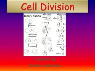
Types of cell division In prokaryotes there is only one simple type of cell division, which produces two identical daughter cells from one progenitor cell (asexual cell division). Eukaryotes also show asexual cell division; this converts a single fertilized egg cell, a zygote, into a multicellular organism, or a single unicellular organism into a population or a colony. The asexual cell division in eukaryotes is called mitosis (M). Both haploid (n) and diploid (2n) cells can divide asexually. The sexual cell division in eukaryotes is called meiosis (Mei) and occur in specialized cells, the meiocytes, which divides twice, resulting in four haploid cells called a tetrad.
Life cycles of humans, plants, and fungi In humans and many plants, three cells of the meiotic tetrad abort. The abbreviation n indicates a haploid cell, 2n a diploid cell; gp stands for gametophyte, the small structure of haploid cells that will produce gametes. In flowering plants the gametophyte stage is radically reduced, but in others (such as mosses) the gametophyte is the main vegetative stage
G1, S, G2, M • Cells spend most of their life in interphase, the period between nuclear divisions, and comparatively little time in mitosis. Interphase is divided into three stages, G1, S, and G2. • In G1cells are growing and synthesizing the materials necessary for their proper functioning. The cells are "doing their thing", so if they are nose cells they are producing mucus, if they are muscle cells they are contracting and relaxing, etc. Some cells, such as our nerve cells (neurons) and red blood cells, never leave this stage and it is then called G0. A cell which is in the G0 stage will not divide. It will not grow, either, but will continue to function until it dies. Cells which will divide pass through a specific phase in G1 which acts as a gateway into the S stage. Once cells pass this "point of no return" they will proceed through S, G2 and mitosis. S is the stage when DNA synthesis(chromosome replication) occurs. The chromosomes consist of two identical strands once replication is completed. Each of these strands is called a chromatid. During mitosis the chromatids will separate and each chromatid will become a separate chromosome.
Mitosis • Mitosis (M) is usually the shortest segment of the cell cycle, lasting for approximately 5 to 10 percent of the cycle. DNA synthesis takes place during the S period. G1 and G2 are gaps between S and M. Together, G1, S, and G2 constitute interphase, the time between mitoses. (Interphase used to be called "resting period"; however, cells are active in many ways during interphase, not the least of which, of course, is DNA replication.) The chromosomes cannot be seen during interphase, mainly because they are in an extended state and are intertwined with one another (chromatine). • For the sake of study, biologists divide mitosis into four stages called prophase, metaphase, anaphase, and telophase. It must be stressed, however, that any nuclear division is a dynamic process on which we impose such arbitrary stages only for our own convenience. • In each of the resultant daughter cells, the chromo-some complement is identical with that of the original cell. Of course, what were referred to as chromatids now take on the role of full-fledged chromosomes in their own right.
The four stages of mitosis • Prophase • Metaphase • Anaphase • Telophase
Prophase • The onset of mitosis is characterized by the chromosomes becoming distinct. They get progressively shorter through a process of contraction, or condensation, into a series of spirals or coils; the coiling produces structures that are more easily moved around. • As the chromosomes become visible, they appear double-stranded, each chromosome being composed of two longitudinal halves, the sister chromatids. The two chromatids formed by one chromosome each contain one of the replicated DNA molecules. • Because of semiconservative replication these replicate DNA molecules are each "half old and half new"; that is, in each double helix one of the nucleotide strands is newly polymerized. These sister chromatids are joined at a region called the centromere. At this stage the centromere has already divided into a pair of sister centromeres. • The nuclear membrane begins to break down, and the nucleoplasm and cytoplasm become one.
Metaphase • At this stage, the nuclear spindle becomes prominent. The spindle is a birdcage-like structure that forms in the nuclear area; it consists of a series of parallel proteinaceous fibers that point to each of two cell poles. These spindle fibers are polymers of a protein called tubulin. The chromosomes move to the equatorial plane of the cell, where one sister centromere becomes attached to a spindle fiber from one pole; the other sister centromere, to the other pole.
Metaphasechromosomes Some of the orders of chromatin packing thoughtto give rise to the highly condensed mitotic chromosome. The folding of naked DNA into nucleosomes is the best understood level of packing. The structures corresponding to the additional layers of chromosome packing are more speculative.
Anaphase and telophase • Anaphase begins when the pairs of sister chromatids separate, one of a pair moving to each pole. The centromeres, which now clearly appear to have divided, separate first. As each chromatid moves, its two arms appear to trail its centromere; a set of V-shaped structures results, with the points of the V's directed at the poles. • At telophase, a nuclear membrane re-forms around each daughter nucleus, the chromosomes uncoil, and the nucleoli reappear, effectively re-forming the interphase nuclei. By the end of telophase, the spindle has dispersed, and the cytoplasm has been divided into two by a new cell membrane.
Cells in active proliferation The apical meristem x400. Most of the cells even in this area of active cell division are at interphase. Those with visible chromosomes are at some stage of mitosis. Longitudinal section of an onion root tip (apical meristem) x40.
Meiosis: Prophase 1 • Meiosis consists of two nuclear divisions, distinguished as meiosis I and meiosis II. The events of meiosis I are quite different from those of meiosis II, and the events of both differ from those of mitosis. Each meiotic division is formally divided into prophase, metaphase, anaphase, and telophase. • PROPHASE I.The chromosomes become visible as long, thin single threads (which are composed of pairs of replicated DNA molecules), and continue contracting during the entire stage. Homologous chromosomes form pairs (this does not happen in Mitosis); each chromosome has a pairing partner, and the two become progressively paired, or synapsed, along their lengths. Thus, the number of homologous pairs of chromosomes in the nucleus is equal to the haploid number n. The beadlike chromomeres align precisely in the paired homologs, producing a distinctive pattern for each pair. Since each member of a homologous pair produces two sister chromatids, the synapsed structure now consists of a bundle of four homologous chromatids, the tetrad. At this stage, cross-shaped structures called chiasmata (singular, chiasma) appear between nonsister chromatids. Each homologous group of four generally has one or more chiasmata. Chiasmata are the visible manifestations of events called crossovers. A crossover is a precise breakage, swapping, and reunion between two nonsister chromatids.
Meiosis: Metaphase I, Anaphase I, and TelophaseI • METAPHASE I.The nuclear membrane and nucleoli disappear, and each pair of homologs takes up a position in the equatorial plane. The sister centromeres do not appear to have divided, so they act as one. This apparent lack of division represents a major difference from mitosis. The two nonsister centromeres attach to spindle fibers from opposite poles. • ANAPHASE I. Homologous chromosomes move directionally to opposite poles. This is the stage at which haploid nuclei are formed. • TELOPHASE I.In many organisms, this stage do not exist, no nuclear membrane re-forms, and the cells proceed directly to meiosis II. In other organisms, telophase I and the interkinesis are brief in duration; the chromosomes elongate and become diffuse, and the nuclear membrane re-forms. In any case, there is never DNA synthesis at this time, and the genetic state of the chromosomes does not change.
Meiosis I Stages of Trillium erectum Late prophase I Metaphase I Anaphase I
Meiosis 2 • PROPHASE II.The presence of the haploid number of sister chromatid pairs in the contracted state characterizes prophase II. • METAPHASE II.The pairs of sister chromatids arrange themselves on the equatorial plane during metaphase II. Here the chromatids often partly dissociate from each other instead of being closely pressed together as they are in mitosis. • ANAPHASE II.Centromeres split and sister chromatids are pulled to opposite poles by the spindle fibers. • TELOPHASE II.The nuclei re-form around the chromosomes at the poles.
Meiosis II Stages of Trillium erectum Metaphase II Anaphase II The centromers have separated, half chromosomes are drawn to the poles Early telophase II. The nuclei are haploid, the chromosomes single-stranded