Download
1 / 18
210 likes | 481 Views
Lab 2. Methods of Leukocyte fractionation: Enrichment: 1. Fluorescence activated cell sorting (FACS) -cost, -availability of equipment -markers may be on more than one cell type e.g. MHC class II on APCs (DC, M, B cells) -markers may not be on a cell type

E N D
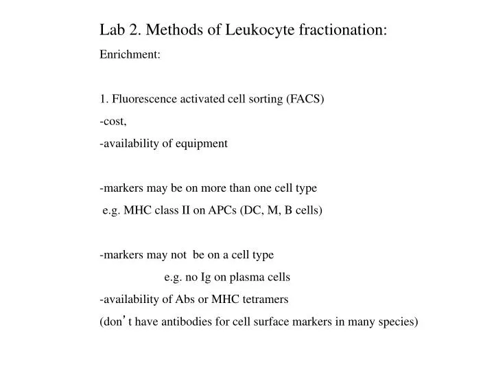
Lab 2. Methods of Leukocyte fractionation: Enrichment: 1. Fluorescence activated cell sorting (FACS) -cost, -availability of equipment -markers may be on more than one cell type e.g. MHC class II on APCs (DC, M, B cells) -markers may not be on a cell type e.g. no Ig on plasma cells -availability of Abs or MHC tetramers (don’t have antibodies for cell surface markers in many species)
1. FACS isolation with specific antibodies or tetramers. Antibody identifies specific cell type (eg. CD3+ T cells) Tetramer of MHC class I with specific peptide for isolation of specific T cells (eg. influenza specific CD8+ T cells)
2. Panning or Magnetic Beads (MACS) for positive or negative selection 2.a Magnetic sorting The magnetically labeled fraction is recovered by applying the vessel to an external magnet. The magnetic fraction can be washed and isolated for subsequent use in downstream procedures, or the supernatant is recovered for further use.
2 b. Cell separation using an AutoMacs magnetic cell sorter The splenocytes are washed (following removal of erythrocytes by osmotic shock) and incubated with anti-CD43 and anti-Mac-1 antibody-conjugated microbeads (Miltenyi Biotec). The bead-bound cells (positive fraction) are separated from unbound cells (negative fraction).
3. Depletion of non-target cells-examples -C’ mediated lysis of a non-target cell- need Abs 4. Enrichment in vivo -e.g. peritoneal priming and lavage 5. Removal of cells adherent to nylon wool. (monocytes stick to nylon wool)
6. Continuous gradient centrifugation for cell separation based -cell fractionation by differential fractionation -density gradient fractionation 7. Discontinuous gradient centrifugation- -granulocytes, RBCs and other dense cells removed -physical characteristics may not distinguish cell types 8. Cytospin to concentrate, stain and visualize cells.
DENSITY DENSITY is the mass per unit volume of a substance, often expressed in g/ml. DIFFERENTIAL CENTRIFUGATION DIFFERENTIAL CENTRIFUGATION separates particles on the basis of their size. By a series of centrifugations at various speeds and times, different-sized particles are sedimented and collected from an initially homogenous suspension. DENSITY GRADIENT CENTRIFUGATION DENSITY GRADIENT CENTRIFUGATION is separation performed in a supporting column of solution in which the density and solution concentration increase toward the bottom of the centrifuge tube. Requires a gradient maker to set up. DISCONTINUOUS, OR STEP, GRADIENT DISCONTINUOUS, OR STEP, GRADIENT is one composed of layers, with abrupt changes in density and/or concentration from one layer to the next.
6. Continuous gradient centrifugation a. Differential centrifugation for fractionation Fractionation of a cell homogenate to isolate nuclei or smaller parts of cell.
6.b. continuous gradient centrifugation -Density gradient centrifugation A gradient maker is used to mix liquids of two densities Close the port between the solutions. Add lower density solution to the output side, higher density to the other. Need rapid mixing in the chamber and make sure you see the higher concentration solution mixing in. Fill centrifuge tubes from the bottom with liquid of increasingly higher density displacing the lower density liquid until the tube is full. Usually a long metal syringe needle is placed in the bottom of the tube so that the density is not really disturbed when the needle is removed.
6. b. Continuous gradient centrifugation low Monocytes Lymphocytes PMNs RBCs spin high To wash isolated cells, resuspend in media and pellet again. Cells are pelleted intact at low speeds 400 x g for 10 minutes
6. b. Continuous gradient separation: Separation on a preparative scale by centrifugation through a sucrose gradient. Two types of hepatitis B capsid Separation and crystallization of T = 3 and T = 4 icosahedral complexes of the hepatitis B virus core protein. Acta Cryst. (1999). D55, 717-720
7. Step Gradient-discontinuous gradient centrifugation Layering blood on Histopaque 1077 serum “buffy coat” or PBL Red blood cells, Platelets, granulocytes Histopaque Unfractionated Blood on Histopaque (note the undisturbed interface) Fractionated blood on Histopaque
7.a. Separation of heparinized blood on Histopaque 1077 -one step Lymphocytes/monocytes Red blood cells, platelets granulocytes.
7.b. Step gradient-multiple steps Percoll density centrifugation for fractionation of B lymphocytes Mature B cells Proliferating B cells Plasma cells Trout cells were layered over step gradients of 50, 60, 70% Percoll. Western blot analysis of Pax-5 and Blimp-1 was used to test the purification. These two transcription factors display distinct and partially overlapping expression patterns during B cell differentiation. mature (resting and early activated) B cells (Pax-5+Blimp–) reside in the 70% layer, proliferating B cells reside in the 60% layer (Pax-5+Blimp+) and plasma cells (Pax-5–Blimp+) reside in the 50% layer. J Immunol. 2005 Jun 1;174(11):6608-16.
8. A cytospin instrument is used to collect cells from a solution for display on a slide. A cytospin device
Centrifugation basics Catastrophic failure of an unbalanced centrifuge Always balance opposing tubes Place test tube in centrifuge holder. The higher the speed the more accurately balanced the centrifuge must be. Never walk away from a Balance with another test tube filled to the same level in the opposite holder.
Centrifuge Basics-continued Make sure you are using the correct rotor for the centrifuge. Never exceed the maximum g force for the rotor. Make sure you are using the correct centrifuge tube for the job. Tubes may break if centrifuged at too high a speed. Even plastic tubes can break if not in appropriate adaptors. Pre-cool centrifuge and rotors. NEVER walk away from a centrifuge until it reaches top speed.
Use of a centrifuge nomogram for conversion of rpm to rcf. Q. You are following a protocol that says: Spin cells at 400 rpm for 10 minutes. What else do you need to know? RCF=11.2 x r (RPM/1000)2 r in cm