Download
1 / 25
250 likes | 325 Views
Explore the complex process of inflammation, its protective role, cardinal signs, functions, and the inflammatory response. Learn about changes in vessel wall permeability, mediators like histamine and bradykinin, and cellular events in inflammation.

E N D
A normal response of living tissues to injury. It prepares the tissue for healing and repair. A dynamic process that lasts from a few minutes to a few years. Depending on: the extent of the injury the type of injury the vascularity of the tissue.
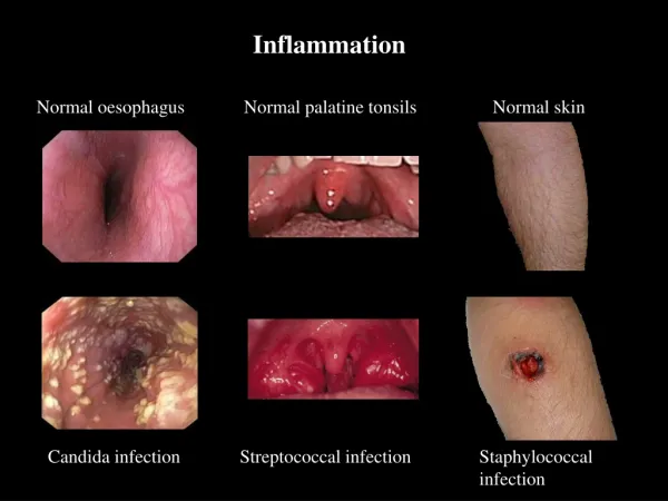
Protective Role • Although inflammation is a necessary process, it must be contained. It serves to inform the individual that an area has been injured. It restricts function to prevent further injury to the area. • Cardinal Signs of Inflammation • Erythema • Heat production • Edema • Pain • Loss of Function.
Functions of Inflammation • Inactivate injurious agent • Break down and remove dead tissue • Initiate healing of tissue.
Inflammatory Response • Complex response that involves: • Circulatory (hemo-dyanamic) changes , changes in vessel wall permeability , response of white blood cells release of soluble mediators. • Smooth muscle cells act as sphincters, regulating the inflow blood into the capillaries. • Relaxation of smooth muscle cells allows blood to rush into the capillaries erythema, edema, and heat.
first response of arterioles to injury… • vasoconstriction (a few seconds in duration). • Followed by… vasodilation • flooding the capillary network with arterial blood • Blood influx into the area dilates capillaries (endothelial cells & basement membrane) blood flow is not effectively regulated.
pressure from the capillaries is transmitted to the venules • have no capacity to contract increased pressure in the capillaries and venules forces: • plasma filtration through the vessel wall • Results in: edema formation.
Dilated Capillaries & Venules • slowed blood flow leads to congestion • sludged, erythrocytes form stacks = rouleaux • impair circulation further. • -Leukocytes (WBC) marginalize become attached to the endothelium “pavementing” • -Leukocytes develop elongated protrusions of the surface cytoplasm, become sticky adhere to the endothelial cells lining the capillaries (particularly those in the postcapillaryvenules.)
WBC adhesion to the surface of the venules • accomplished by surface adhesion molecules , normally present on leukocytes and endothelial cells in an inactive form during inflammation, activated by soluble mediators of inflammation interleukins,a type of cytokine (protein) derived from platelets and leukocytes, Normally present in blood in small amounts Concentrations increase at site of inflammation , Mediate communication among leukocytes and other cells active in inflammation or cell mediated immune response, The result is a maximized response to a microorganism or other foreign antigen
Changes in Vessel Wall Permeability • Occurs in capillaries and postcapillaryvenules ,Changes occur due to increased pressure inside the blood vessels slowed circulation , reduction in the oxygen supply and nutrients to endothelial cells adhesion of leukocytes and platelets to endothelial cells release of soluble mediators of inflammation from inflammatory cells
Mediators of Inflammation • - Chemical Mediators • Plasma derived circulate in an inactive form must be transformed into an active form by an activator numerous, specific and non-specific ,all activators have natural in-activators to maintain balance • Cell derived may be pre-formed and stored in granules of platelets and leukocytes (histamine)…or… may be synthesized as needed.
effects include • vasodilation • vasoconstriction • altered vascular permeability • activation of inflammatory cells • chemotaxis • cytotoxicity • degradation of tissue • pain • fever.
Mediators of Inflammation • Biogenic amines • Histamine • Peptides : Bradykinin ,Complement system • histamine , released from mast cells, basophils and platelets cause a contraction of the endothelial cells of venules ,gaps form, increasing vessel permeability , fluids and blood cells exit into interstitial space ,effect rapidly inactivated by histaminases. • bradykinin . Plasma protein formed through the activation of a coagulation factor (XII) leads to the activation of several biological systems. • Arachadonic Acid Derivatives , derived from the phospholipids of cell membranes ,Involved in all stages of inflammation. • cyclooxygenase pathway (COX) ,formation of prostaglandins, Modulate vasomotor tone ,modulate platelet aggregation and thrombosis , promote pain perception and mediate fever.
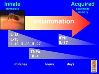
Cellular Events in Inflammation • Increased permeability of the vessel walls of postcapillaryvenules and capillaries.... leakage of fluid from the vessels into the interstitial spaces. “transudation” edema formation. • Polymorphonuclear Leukocytes • 60-70% of all WBCs , 2-4 day lifespan, first to appear in acute inflammation ,highly mobile ,bacteriocidally active perform phagocytosis , produce and release mediators of inflammation cytokenes interleukin-1.....a pyrogen that acts on the hypothalamus......causes fever.
as inflammation evolves, PMNs are joined by monocytes and eosinophils (within 48 hrs) as inflammation progresses into chronic phase, PMNs are replaced by macrophages, lymphocytes, and plasmacells. • - adhesion of PMNs to the endothelial cells, insertion of cytoplasmic pseudopods between the junctions of endothelial cells, passage through the basement membrane, ameboid movement away from the vessel toward the cause of inflammation (chemotaxis).
The Inflammatory Process • Phagocytosis • PMNs reach the bacteria or irritant lose mobility begin acting as scavengers active uptake of bacteria or other cellular debris lysosomal degranulation of irritant PMNs die in the process released in a yellow viscous fluid is pus (purulent).
Cells of Inflammation • -Eosinophils • 2-5% of WBCs appear 2-3 days after the PMNs, slower to react, slower mobility ,single nucleus , prominent in allergic reactions, hay fever, asthma ,parasitic infections ,live longer than PMNs, are present in chronic inflammation
-Basophils • less than 1% of WBCs most prominent in allergic reactions regulated by immunoglobulin E, rich in vasoactive substances, histamine, precursors of mast cells. • -Macrophages • tissue cells (histocytes) • appear 3-4 days after infection or tissue destruction ,long lifespan, present in chronic inflammation • capable of phagocytosis rich in lytic enzymes, secrete cytokines locally and systemically, recruit lymphocytes to site of inflammation • produce lymphocyte growth factors and fibroblast growth factors, arachadonic acid metabolites activate, coagulation sequence and thrombolysis.
lymphocytes • main means of providing the body with immunity ,20-40% of the WBCs activated by the presence of a specific antigen. • -platelets fragments of cytoplasm released from bone marrow no nucleus , cytoplasm contains vacuoles with 3 types of granules.
Classification of Inflammation in Clinical Practice • Classification based on • duration • etiology • location • morphology or pathological characteristics
on duration • “Acute Inflammation” lasts from a few hours to a few days , Sudden onset, short duration, severe symptoms (possesses the cardinal signs of inflammation heat, erythema, edema, pain, loss of function) • Recurrent- acute inflammation that occurs in bouts • Chronic Inflammation : Represents: extension of an acute inflammation , prolonged healing of an acute inflammation persistence of causative agents or May evolve without acute • chronic inflammation may develop in response to a foreign substance, foreign body granulomas develop around a objects in subcutaneous tissues. • Acute PMNs regulated • Chronic :Macrophages, lymphocytes, and plasma cells ***Much too simplistic*
Etiology • “The study of the causes of disease.” • Inflammations are caused by • infectious pathogens bacteria , viruses ,protozoans, fungi , and helminthic origins (wormlike animals) • chemical 1organic /inorganic 2 industrial /medicinal 3 exogenous /endogenous • physical1 foreign bodies 2 heat 3irradiation 4trauma • immune factors
On Location • Localized • Widespread or Systemic, involving multiple organs , bacteria spread throughout the blood stream • Pathological Characteristics • -Serous , clear exudate (blisters) , acute inflammatory response , Mildest form of inflammation. • -Fibrinous , exudate rich in fibrin , Relatively severe inflammation , Common with bacterial infections • -Purulent ,pus formation Can occur on mucosa, skin…or In internal organs