Download
1 / 23
230 likes | 527 Views
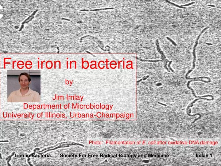
Free iron in bacteria by Jim Imlay Department of Microbiology University of Illinois, Urbana-Champaign. Photo: Filamentation of E. coli after oxidative DNA damage. Iron in Bacteria Society For Free Radical Biology and Medicine Imlay 1. The problem with intracellular "free" iron.

E N D
Free iron in bacteria by Jim ImlayDepartment of MicrobiologyUniversity of Illinois, Urbana-Champaign Photo: Filamentation of E. coli after oxidative DNA damage Iron in Bacteria Society For Free Radical Biology and Medicine Imlay 1 Iron in BacteriaSociety For Free Radical Biology and Medicine Imlay 1
The problem with intracellular "free" iron Most biological molecules cannot be damaged at a significant rate by direct reactions with molecular oxygen, superoxide, or hydrogen peroxide. However, they can be oxidized by hydroxyl radical (HO•). This species is formed when a single electron is transferred to H2O2 e- + H2O2 ---> HO• + OH- In in vitro systems the most facile donors of single electrons to H2O2 are transition metals, most notably iron (II) and copper (I). Fe2+ + H2O2 ----> HO• + OH- + Fe3+ (the Fenton reaction [1]) Cu1+ + H2O2 ----> HO• + OH- + Cu2+ Although organic electron donors, such as reduced quinones, are not thermodynamically prohibited from transferring electrons to H2O2, they are kinetically limited. No examples of such "organic Fenton reactions" have yet withstood scrutiny (but see [2]). Therefore, the vulnerability of intracellular DNA and proteins to oxidation should depend in part upon the concentration of available iron and copper. Iron in Bacteria Society For Free Radical Biology and Medicine Imlay 2
Iron catalyzes HO• formation in vivo (1) xth-1 + dipyridyl 1 • Although either copper or iron suffices for H2O2 reduction in vitro, iron is the responsible species in vivo. • There are three primary pieces of evidence that support this conclusion: • First, iron chelators that can penetrate bacteria--dipyridyl, o-phenanthroline, and desferrioxamine--prevent exogenous H2O2 from damaging DNA [3]. In this figure dipyridyl fully prevents H2O2 from killing a strain of E. coli that cannot repair oxidative DNA lesions. The same result is obtained from direct measurements of DNA lesions. 0.1 xth-1 0.01 0.001 0 5 10 15 Minutes exposure to 0.75 mM H2O2 Iron in Bacteria Society For Free Radical Biology and Medicine Imlay 3
Iron catalyzes HO• formation in vivo (2) Second, the kinetics with which H2O2 damages intracellular DNA indicate the mediation of a ferryl radical (FeO2+). Ferryl radicals are the immediate products of electron transfer from Fe2+ to H2O2 [4]; they subsequently dissociate to form Fe3+ + HO•: Fe2+ + H2O2 ----> [FeO2+] + OH- + H+ ----> Fe3+ + HO• High concentrations of H2O2 can scavenge ferryl radicals before HO• is formed: FeO2+ + H2O2 ----> Fe3+ + H2O + O2•- DNA damage is therefore actually more abundant at lower concentrations of H2O2 (left graph, next slide). This is not true of the copper-mediated reaction (not shown). When intact cells are exposed to H2O2 , high concentrations of H2O2 again suppress the rate of DNA damage (right graph, next slide), thus indicating that HO• formation inside cells is mediated by iron rather than copper [5]. Iron in Bacteria Society For Free Radical Biology and Medicine Imlay 4
[2, cont’d] Damage suppression by excess H2O2 indicates the mediation of FeO2+ 1.5 1 xth-1 mutant was exposed to indicated [H2O2] for 15 minutes. Lesions created in circular DNA exposed in vitro to 80 nM ferrous iron + indicated [H2O2]. 0.1 1 0.01 0.5 0.001 0.0001 0 0 1 2 3 4 0 2.5 5 7.5 10 Iron in Bacteria Society For Free Radical Biology and Medicine Imlay 5
Iron catalyzes HO• formation in vivo (3) • Third, E. coli mutants that over-import iron are unusually vulnerable to DNA damage by exogenous H2O2 [6, 7]. Overexpression of ferritin, a storage protein that specifically sequesters iron, prevents damage [6]. Why doesn't copper contribute to HO• formation in vivo? The amount of available copper may be too small. However, even mutants that have lost the ability to control copper levels exhibit normal resistance to H2O2 [8]. Thus, a second factor may be that copper is liganded by the large pool of intracellular thiols, including glutathione. Millimolar levels of glutathione block the participation of copper in HO• formation in vitro [9]. Iron in Bacteria Society For Free Radical Biology and Medicine Imlay 6
“Free” iron is the source of toxic hydroxyl radicals Most iron inside cells is stably incorporated into proteins. Some of this iron is solvent-exposed and can be oxidized by H2O2. This is true, for example, of dehydratases that contain surface-exposed iron-sulfur clusters [10]. It follows that the HO• that is formed by this reaction could potentially oxidize the side chains of these iron-binding proteins. However, the immense reactivity of HO• precludes the possibility that it will diffuse far from the site of its formation before it reacts with a biomolecule or metabolite. Thus, protein-integrated iron is unlikely to generate the HO• that damages DNA. These considerations suggest that the iron that catalyzes DNA damage is likely to be adventitiously localized on the surface of DNA or bound to small metabolites that can diffuse close to the DNA [11]. This iron is commonly denoted "free iron," to indicate that it is not integrated into enzymes. • The term "free iron" is not intended to suggest that the iron is hexa-aqueous. Iron binds avidly to virtually all biomolecules, so iron atoms free within the cell are likely to adhere to the surfaces of membranes, nucleic acids, proteins, etc. Iron in Bacteria Society For Free Radical Biology and Medicine Imlay 7
Total metal analyses can quantify the amount of iron inside cells, but most of that iron is stably incorporated into proteins and is uninvolved in Fenton chemistry. To focus specifically upon free iron, either Mossbauer [12] or epr [13] methods are preferable. An important advantage of these methods is that they can be applied to whole cells. EPR is the more convenient of the two techniques. This method most easily detects ferric iron. (Ferrous iron is also epr-active but it displays a broad, indistinct signal.) Recent studies indicate that most of the free iron in E. coli is in the reduced form and therefore relatively invisible to epr [14]. However, the iron can be oxidized by treatment of the cells with either H2O2 or desferrioxamine. The latter agent binds and lowers the reduction potential of iron, triggering its autoxidation and trapping it in the ferric state. Thus exposure of E. coli to either of these agents allows the free iron pool to be visualized. Intracellular free iron can be quantified Iron in Bacteria Society For Free Radical Biology and Medicine Imlay 8
Free iron levels in E. coli g = 4.3 • Such experiments indicate that growing E. coli cells contain 15-30 micromolar free iron [13]. In such an experiment, the functional definition of "free iron" is iron that is redox-active and that it can be chelated by desferrioxamine. Since desferrioxamine blocks DNA damage, this includes the iron the catalyzes DNA damage. • The Fur mutants that have unregulated iron uptake, and that are highly vulnerable to DNA damage, contain approximately 90 micromolar free iron [13]. Ferric iron standard Wild-type cells: ca. 20 uM iron Fur mutant: 85 uM iron EPR spectra of Fe(III)desferrioxamine, i.e. ferrioximine. With a typical x-band epr these signals are centered at about 1550 gauss; Hpp 50 G. Iron in Bacteria Society For Free Radical Biology and Medicine Imlay 9
Why is there free iron in the cell? • In no organism do we understand the route by which iron is trafficked from transport-complexes to its ultimate destination in metalloproteins. However, it seems inconceivable that iron is merely dumped into the cell upon import -- not only because free iron catalyzes oxidative damage, but also because iron sticks so avidly to biomolecules that it might never find its way to the target proteins. This is an important gap in our understanding of iron metabolism. • This argument implies the existence of a pipeline of iron flow from transporter to incorporation. If this is correct, then free iron represents iron that has escaped the usual pathways of iron traffic. Iron in Bacteria Society For Free Radical Biology and Medicine Imlay 10
Oxidants release iron from some [4Fe-4S] clusters cys S Fe cys Fe S Fe S cys S Fe H2O2 O2- Fe2+ • One mechanism of escape is through disintegration of protein iron-sulfur clusters. In particular, clusters in dehydratases fall apart when they are oxidized into an unstable valence [15-19]: Protein-[4Fe-4S]2+ + O2•- + 2H+ ----> Protein-[4Fe-4S]3+ + H2O2 Protein-[4Fe-4S]3+ ---> Protein-[3Fe-4S]1+ + Fe2+ • Superoxide is a particularly good oxidant (right); the rate constant for this reaction is ca. 106 m-1 s-1[10]. Peroxynitrite also rapidly destabilizes the clusters of these enzymes [20, 21]. H2O2 itself does so, but more sluggishly [10]. Iron in Bacteria Society For Free Radical Biology and Medicine Imlay 11
Free iron during oxidative stress • The level of free iron is elevated when E. coli is exposed to redox-cycling drugs that generate superoxide. Mutants that lack SOD contain almost 10 times the amount of free iron as do wild-type cells [13]. For this reason, HO• is formed at a proportionately higher rate when these cells are exposed to H2O2 [22]. Rapid DNA damage ensues (next slide). Wild-type g = 4.3 Wild-type + paraquat SOD-deficient mutant (EPR spectra of ferrioximine are shown at equivalent gain.) Iron in Bacteria Society For Free Radical Biology and Medicine Imlay 12
The abundant free iron in superoxide-stressed cells increases their vulnerability to DNA damage 1 Wild-type SOD-deficient mutant 0.1- 0 5 10 15 Minutes of H2O2 Exposure Iron in Bacteria Society For Free Radical Biology and Medicine Imlay 13
Free iron without oxidative stress • However, the free iron found in wild-type (SOD-proficient) cells does not arise from superoxide-mediated damage, since SOD overproduction cannot further diminish either the epr-detectable free-iron signal or the rate of HO• formation. In fact, the amount of free iron is actually higher in anaerobic than aerobic E. coli, as the Feo iron-transport system is induced [7]. • This basal free iron may arise from spontaneous iron leakage from dehydratases or other proteins. Alternatively, iron may be trafficked by a weak chelator that does not preclude either its detection by epr or its participation in Fenton chemistry. It is notable that both Fur and aconitase B, two proteins that control iron acquisition, appear to bind iron reversibly, as if their regulatory action depends on the equilibration of iron between them AND an accessible iron pool in the cell [23, 24]. The nature of that pool remains unknown. Iron in Bacteria Society For Free Radical Biology and Medicine Imlay 14
How does the cell control the amount of free iron? • Given the role of free iron in creating DNA damage, it is unsurprising that bacteria have evolved methods to scavenge it. Experiments in which cells were exposed to a bolus of peroxynitrite revealed that free-iron levels rose and then fell within a minute [25]. The disappearance of the free iron exceeded the pace at which the damaged iron-sulfur clusters were repaired, suggesting that the free iron was scavenged. Iron in Bacteria Society For Free Radical Biology and Medicine Imlay 15
Is Dps an iron scavenger? • E. coli synthesizes three proteins--ferritin [26], bacterioferritin [27], and Dps [28] ; each sequester many atoms of iron. Ferritin and bacterioferritin are synthesized when iron is highly available in the environment, and thus they appear to be the routine storehouses of iron. They presumably donate the stored iron to metallation processes when iron becomes scarce. Consistent with this idea, mutants that lack these proteins cease growth more rapidly than wild-type cells when iron-starvation is imposed [29]. • In contrast, Dps is induced by the OxyR regulatory protein specifically in response to the presence of H2O2 [30]. Mutants that lack Dps are particularly sensitive to oxidative DNA damage [31]. In vitro this protein can both store iron and bind to DNA. Its protective role in vivo may be stem from a combination of these activities. Iron in Bacteria Society For Free Radical Biology and Medicine Imlay 16
Outlook • The chemistry of oxidative damage: iron leakage from oxidized dehydratases and its participation in Fenton chemistry is likely to be the same in all organisms. These processes, for example, have also been observed in yeast and in mammalian cells [32-35]. It is notable, though, that several bacteria have few (or no) iron enzymes and therefore may be exempt from this kind of damage [36, 27]. The vulnerability of still other organisms to H2O2 varies widely [38], perhaps reflecting differences in their free-iron content. • Future work: despite the sophisticated biochemical and genetic strategies that can be brought to bear upon bacteria, we still know remarkably little about the physical mechanisms of iron transport, storage, and regulation, and virtually nothing about iron trafficking and its insertion into metalloproteins. These areas are ripe for future work. Iron in Bacteria Society For Free Radical Biology and Medicine Imlay 17
References 1. Walling, C. 1975. Fenton's reagent revisited. Accounts of Chemical Research8:125-131. 2. Zhu, B.-Z., H.-T. Zhao, B. Kalyanaraman, and B. Frei. 2002. Metal-independent production of hydroxyl radicals by halogenates quinones and hydrogen peroxide: an ESR spin trapping study. Free Rad. Biol. Med.32:465-473. 3. Imlay, J. A. and S. Linn. 1988. Toxic DNA damage by hydrogen peroxide through the Fenton reaction in vivo and in vitro. Science240:640-642. 4. Rush, J. D., Z. Maskos, and W. H. Koppenol. 1990. Distinction between hydroxyl radical and ferryl species. In: Methods in Enzymology186:148. 5. Imlay, J. A. and S. Linn. 1986. Bimodal pattern of killing of DNA-repair-defective or anoxically grown Escherichia coli by hydrogen peroxide. J. Bacteriol.166:519-527. 6. Touati, D., M. Jacques, B. Tardat, L. Bouchard, and S. Despied. 1995. Lethal oxidative damage and mutagenesis are generated by iron in Dfur mutants of Escherichia coli: protective role of superoxide dismutase. J. Bacteriol.177:2305-2314. Iron in Bacteria Society For Free Radical Biology and Medicine Imlay 18
References (cont’d) 7. Keyer, K., A. S. Gort, and J. A. Imlay. 1995. Superoxide and the production of oxidative DNA damage. J. Bacteriol.177:6782-6790. 8. Brauer and Imlay, unpublished data. 9. Imlay and Linn, unpublished data. 10. Flint, D. H., J. F. Tuminello, and M. H. Emptage. 1993. The inactivation of Fe-S cluster containing hydro-lyases by superoxide. J. Biol. Chem.268:22369-22376. 11. Luo, Y., Z. Han, S. M. Chin, and S. Linn. 1994. Three chemically distinct types of oxidants formed by iron-mediated Fenton reactions in the presence of DNA. Proc. Natl. Acad. Sci. USA 91:12438-12442. 12. Khoroshilova, N., C. Popescu, E. Munck, H. Beinert, and P. Kiley. 1997. Iron-sulfur cluster disassembly in the FNR protein of Escherichia coli by O2: [4Fe-4S] to [2Fe-2S] conversion with loss of biological activity. Proc. Natl. Acad. Sci. USA 94:6087-6092. 13. Keyer, K. and J. A. Imlay. 1996. Superoxide accelerates DNA damage by elevating free-iron levels. Proc. Natl. Acad. Sci. USA 93:13635-13640. Iron in Bacteria Society For Free Radical Biology and Medicine Imlay 19
References (cont’d) 14. Woodmansee, A. N. and J. A. Imlay. 2002. Reduced flavins promote oxidative DNA damage in non-respiring E. coli by delivering electrons to intracellular free iron. J. Biol. Chem., in press. 15. Kuo, C. F., T. Mashino, and I. Fridovich. 1987. a,b-dihydroxyisovalerate dehydratase: a superoxide-sensitive enzyme. J. Biol. Chem.262:4724-4727. 16. Gardner, P. R. and I. Fridovich. 1991. Superoxide sensitivity of the Escherichia coli 6-phosphogluconate dehydratase. J. Biol. Chem.266:1478-1483. 17. Gardner, P. R. and I. Fridovich. 1991. Superoxide sensitivity of the Escherichia coli aconitase. J. Biol. Chem.266:19328-19333. 18. Liochev, S. I. and I. Fridovich. 1993. Modulation of the fumarases of Escherichia coli in response to oxidative stress. Arch. Biochem. Biophys.301:379-384. 19. Flint, D. H. and R. M. Allen. 1996. Iron-sulfur proteins with nonredox functions. Chem. Rev.96:2315-2334. 20. Castro, L., M. Rodriquez, and R. Radi. 1994. Aconitase is readily inactivated by peroxynitrite, but not by its precusor, nitric oxide. J. Biol. Chem.269:29409-29415. Iron in Bacteria Society For Free Radical Biology and Medicine Imlay 20
References (cont’d) 21.Hausladen, A. and I. Fridovich. 1994. Superoxide and peroxynitrite inactivate aconitases, but nitric oxide does not. J. Biol. Chem.269:29405-29408. 22.McCormick, M. L., G. R. Buettner, and B. E. Britigan. 1998. Endogenous superoxide dismutase levels regulate iron-dependent hydroxyl radical formationin Escherichia coli exposed to hydrogen peroxide. J. Bacteriol.180:622-625. 23. Bagg, A. and J. B. Neilands. 1987. Ferric uptake regulation protein acts as a repressor, employing iron(II) as a cofactor to bind the operator of an iron transport operon in Escherichia coli. Biochemistry26:5471-5477. 24. Varghese and Imlay, manuscript submitted 25. Keyer, K. and J. A. Imlay. 1997. Inactivation of dehydratase [4Fe-4S] clusters and disruption of iron homeostasis upon cell exposure to peroxynitrite. J. Biol. Chem.272:27652-27659. 26. Hudson, S. J., S. C. Andrews, C. Hawkins, J. M. Williams, M. Izuhara, F. C. Meldrum, S. Mann, P. M. Harrison, and J. R. Guest. 1993. Overproduction, purification and characterization of the Escherichia coli ferritin. Eur J Biochem218:985-995. Iron in Bacteria Society For Free Radical Biology and Medicine Imlay 21
References (cont’d) 27. Yariv, J., A. J. Kalb, R. Sperling, E. R. Bauminger, S. G. Cohen, and S. Ofer. 1981. The composition and the structure of bacterioferritin of E. coli. Biochem J.197:171-175. 28. Zhao, G., P. Ceci, A. Ilari, L. Giangiacomo, T. M. Laue, E. Chiancone, and N. D. Chasteen. 2002. Iron and hydrogen peroxide detoxification properties of DNA-binding protein from starved cells. A ferritin-like DNA-binding protein of Escherichia coli. J. Biol. Chem.277:27689-27696. 29. Abdul-Tehrani, H., A. J. Hudson, Y. S. Chang, A. R. Timms, C. Hawkins, J. M. Williams, P. M. Harrison, J. R. Guest, and S. C. Andrews. 1999. Ferritin mutants of Escherichia coli are iron deficient and growth imparied, and fur mutants are iron deficient. J. Bacteriol.181:1415-1428. 30. Altuvia, S., M. Almiron, G. Huisman, R. Kolter, and G. Storz. 1994. The dps promoter is activated by OxyR during growth and by IHF and sigma S in stationary phase. Mol. Microbiol. 13:265-272. 31. Martinez, A. and R. Kolter. 1997. Protection of DNA during oxidative stress by the nonspecific DNA-binding protein Dps. J. Bacteriol.179:5188-5194. Iron in Bacteria Society For Free Radical Biology and Medicine Imlay 22
References (cont’d) 32. Gardner, P. R., I. Raineri, L. B. Epstein, and C. W. White. 1995. Superoxide radical and iron modulate aconitase activity in mammalian cells. J. Biol. Chem.270:13399-13405. 33. Longo, V. D., L.-L. Liou, J. S. Valentine, and E. B. Gralla. 1999. Mitochondrial superoxide decreases yeast survival in stationary phase. Arch. Biochem. Biophys.365:131-142. 34. Srinivasan, C., A. Liba, J. A. Imlay, J. S. Velaentine, and E. B. Gralla. 2000. Yeast lacking superoxide dismutase(s) show elevated levels of "free iron" as measured by whole cell electron paramagnetic resonance. J. Biol. Chem.275:29187-29192. 35. Meneghini, R. 1997. Iron homeostasis, oxidative stress, and DNA damage. Free Rad. Biol. Med.23:783-792. 36. Archibald, F. 1983. Lactobacillus plantarum, an organism not requiring iron. FEMS Microbiology Letters19:29-32. 37. Posey, J. E. and F. C. Gherardini. 2000. Lack of a role for iron in the Lyme disease pathogen. Science288:1651-1653. 38. Pericone, C. D., D. Bae, M. Shchepetov, T. McCool, and J. N. Weiser. 2002. Short-sequence tandem and nontandem DNA repeats and endogenous hydrogen peroxide production contribute to genetic instability of Streptococcus pneumoniae. J. Bacteriol.184:4392-4399. Iron in Bacteria Society For Free Radical Biology and Medicine Imlay 23