Download
1 / 62
901 likes | 2.11k Views
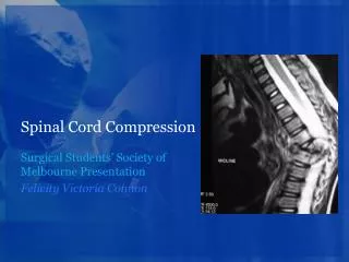
Postgraduate seminar Neurology Unit By: Chidimma A Onwurah. Spinal Cord Compression. Outline . Introduction Anatomy Aetiology Epidemiology Clinical features Investigations Management Conclusion . Spinal Cord Function.

E N D
Postgraduate seminar Neurology Unit By: Chidimma A Onwurah Spinal Cord Compression
Outline • Introduction • Anatomy • Aetiology • Epidemiology • Clinical features • Investigations • Management • Conclusion
Spinal Cord Function • Transmits neural signals and contains neural circuits that control reflexes • Four major functions: • Motor • Sensory • Reflex • Autonomic function
Anatomy • Spinal cord terminates at L1-L2 • Covered by 3 connective tissue: Dura, Pia & Arachnoid • Dural sac ends at S2 • Terminology • Conusmedullaris: most distal bulbous part • Filumtermiale: tapering part of conusmedullaris (mostly fibrous tissue) • Caudaequina: distal collection of nerve roots
3 layers of spinal coverings • Pia mater • Arachnoid mater • Dura mater
Gray matter of spinal cord Cell bodies of neurons which receive afferent information from spinal nerves and send it toward the brain Cell bodies of neurons which receive efferent information from the brain and send it to smooth myocytes, cardiac myocytes, and glands (autonomic motor innervation) Cell bodies of neurons which receive efferent information from the brain and send it to skeletal myocytes (somatic motor innervation)
White matter Carries afferent information into dorsal horn of gray matter Carries efferent information away from ventral horn of gray matter
Arterial supply to the spinal cord • Intraspinal Arteries • Anterior spinal artery • Supply the ventral gray and white matter, except the dorsal horns and dorsal white matter • Posterior spinal arteries • Supply the dorsal horns and dorsal white matter • Extraspinal Arteries • Radicular arteries
Causes of Spinal Cord Compression • Extradural • Vertebral: epidural abscess /Infection- TB & bacterial; vertebral collapse, disc prolapse, spondylosis, spondylolisthesis, Paget’s dx, neoplasm (10 & 20) • Non- vertebral: Hodgkin’s leukemic infiltration or metastases, abscess, cyst
Contd • INTRADURAL • A] Extramedullary • Neoplasm, nerve sheath tumours (schwannomas and neurofibromas ) meningioma; arachnoiditis; arachnoidcyst • B] Intramedullary • Neoplasm, astrocytomas, ependymomas, hemangioblastomas (primary spinal tumours); cyst/syringomyelia; haematomyelia [AVMalformation] and venous angiomas
Epidemiology • USA: spinal stenosis (54%), tumors (26%), ischemia (8%), infection (7%) • Gupta et al in India(2005-2008): spinal tumors (27%), TB spine (25%), transverse myelitis (22%) • Ogunniyi et al in Ibadan (1988-1993): spondyloticmyelopathy (30%), TB spine (25%), neoplastic (12%)
History (SCCx) • Onset • Deficits • Motor • Sensory • Progression • Time to maximum disability • Changes in deficits • Sphincter dysfunction • Autonomic changes • Preceding illness • Systemic complaints
Clinical findings • UMN: preserved muscle bulk or atrophy of disuse, no fibrillation/ fasciculation, hypertonia, brisk DTR, extensor plantar response, absent abdominal reflex • These occur late with intramedullary lesions but early with extramedullary lesion. • LMN: wasting, fibrillation/fasciculation, hypotonia, hyporeflexia, flexor plantar response • Inverted supinator jerk : C5/C6 lesion
Lasegue’s sign – lifting a straight lower limb causes pain. Shows a herniated disc • Shober’s sign- <5cm increase in distance between standing and bending to touch the toes when marks are placed 10cm above and 5 cm below the L5 vertebra Positive in ankylosingspondylosis
Syringomyelia • Fluid filled cavitation in the center of the cord • May be late sequelae of trauma • Most common site is the cervical cord • Loss of pain and temperature • Cape sensory loss • Weakness of muscles in arms with atrophy and hyporeflexia • Later- leg weakness, spasticity with brisk reflexes
Syringomyelia • Can be associated with Arnold Chiari malformation, spinal arachnoiditis, scoliosis, spinal vertebrae misalignment, spinal tumors, spinabifida, meningitis • Other central cord lesions: • Hydromyelia, Hematomyelia • Intramedullary tumors
Cervical spondylosis with myelopathy • Most common form of myelopathy • Degenerative disease of the spine usually at mid and lower cervical spine vertebrae • Narrowing of spinal canal and vertebral foramina • Progressive injury of the spinal cord and spinal roots • Prevalence of cervical degenerative disease reaches 95 % at the age of 65 years
Pathology • Fraying of annulus fibrosus • Extension of disc material in the spinal canal • Bulging of annulus fibrosus • Osteophytes • Hypertrophied longitudinal ligaments and ligamentum flavum • Compression of posterior roots may lead to degeneration in posterior columns • Compression of spinal cord may produce demyelination or focal necrosis
Pathogenesis • No clear explanation • Compression • High mobility of lower cervical vertebrae • Diminished AP diameter of the spinal canal • Compression of spinal arteries and ischemia • Trauma from sudden extreme extension
Clinical features • Radiculopathy • Spondylotic Myelopathy • Myeloradiculopathy • Rotational vertebral artery occlusion, Vertebro-basillar insufficiency • Lhermitte’s • Spurling’s- turning the patients head to the affected side and flexing the neck leads to pain along the affected nerve/dermatome. • Intermittent neurogenic claudication
Bladder disturbance • Fingers may feel swollen or clumsy • Weakness of small muscles of the hand • Clumsiness • Weakness of the legs • Spasticity is more than weakness • Quadriparesis
Muscle stretch reflexes • Loss of biceps reflex, but exaggerated triceps reflex • Extensor plantar reflex • Sensory Changes • Impaired vibration sense, joint position sense • Romberg sign may be present • Sensory gait ataxia in some subjects • Radiculopathy • Symptoms usually begin on waking • Symptoms worsen with Valsalva activities • Motor and reflex changes • C5/C6 disc herniation compresses C6 root • C6/C7 disc herniation compresses C7 root
Brown-sequard syndrome/lateral cord syndrome • Cord hemisection • Trauma or tumour • De-myelination • Burst fracture • Sign and symptoms • Weakness on side of lesion • Dissociated sensory loss • Contralateral loss of pain and temperature to lesion, 1 or 2 levels below; • Ipsilateralpropioceptive loss 1-2 segments below
Posterior cord syndrome • Causes: • Cervical spondylosis, • Multiple sclerosis • Demyelination • Spinal cord tumor • Atlanto-axial subluxation • Sub-acute combined degeneration • Signs • Sensory ataxia (propioceptive loss) • Urinary incontinence • Lhermitte signs
Vascular disease of cord • Arteriovascular malformation and venous angiomas • Occur primarily in the thoracic cord • May present as acute, subacute or chronic compressive lesion • Can cause recurrent symptoms • Associated with pain and bloody CSF if they bleed.
Complete cord • Trauma- commonest cause • Features: • Loss of sensory • Loss of motor • Loss of autonomic • All below the level of lesion
Conusmedullaris syndrome D12 burst fracture compresses the conus. All lumbar and sacral segments can be compressed
Conus medullaris syndrome • Early disturbance of bladder and bowel control, urine retention, constipation • Erectile impotence • Symmetrical saddle anesthesia • Pain is not common, but may occur late
CAUDA EQUINA SYNDROME Acute central disc prolapse L4/5. Medially placed sacral roots sustain maximum compression
Cauda equina syndrome • Early radicular pain in the distribution of lumbosacral roots, usually asymmetric • Pain • May be unilateral • Worse when lying down • Flaccid paresis - glutei, posterior thigh muscles, anterolateral leg muscles, and foot • Asymmetric saddle anesthesia • Loss of ankle jerk • Sphincter disturbance uncommon
Spinal Tuberculosis • Dorsolumbar region is the most frequently involved with D.11 vertebra being most often affected • Radiological features: • disc space narrowing only • kissing lesions; half-moon • wedge collapse of vertebra; • vertebra plana; • lesions localised in the vertebral body and/or its appendages • para-spinal abscesses; • complete destruction of vertebral body.
Other causes of spinal canal narrowing • Ankylosing spondylitis • Ossification of posterior longitudinal ligament • Paget disease • Achondroplasia • Platybasia and basilar invagination
Intra spinal tumors • Most intraspinal tumours are benign • Effects are usually due to compression rather than infiltration • Anatomical groups • Intramedullary • Primary • Metastasis • Extramedullary • Primary • Metastasis
Primary neoplasms • Intramedullary • Astrocytomas, oligodendrogliomas, Ependymomas lipomas, teratomas • Extramedullary • Neurofibromas, more intradural than extradural • Meningiomas, sarcomas, vascular tumours, chordomas
Secondary neoplasms • Extramedullary • Extradural • Carcinomas, lymphoma, myeloma • Intradural • Usually lymphoma spreading to the meninges • Intramedullary • Bronchogenic carcinoma
Clinical features: tumors • Compression of spinal cord by tumor reduces the CSF space arround the cord • Loculation of CSF below the lesion • Increase protein and xanthochromia (Froin Syndrome
Foramen magnum syndrome • Pain • Suboccipital • Lhermitte symptoms • Quadriparesis • Neck stiffness • Atrophy of the muscles of the hands and dorsal neck muscles • Lower cranial nerve palsies, IX – XII • Horner’s syndrome • Papilledema • Downbeat nystagmus • Cerebellar ataxia
Common Investigation • Spinal Xray- plain and contrast • Spinal CT • Spinal MRI • Bone scan • Tumor markers