Download
1 / 24
260 likes | 583 Views
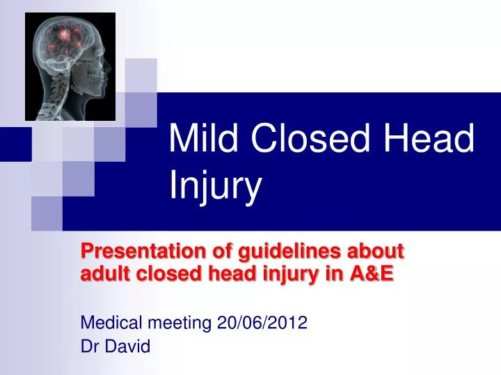
Mild Closed Head Injury. Presentation of guidelines about adult closed head injury in A&E Medical meeting 20/06/2012 Dr David. Case Report HN 329949. Enfant de 1 an et 7 mois (19 months child)

E N D
Mild Closed Head Injury Presentation of guidelines about adult closed head injury in A&E Medical meeting 20/06/2012 Dr David
Case Report HN 329949 • Enfant de 1 an et 7 mois (19 months child) • Il y a 3 heures, elle a ete victime d’un accident de route avec une moto. Sa tête heurte contre la terre. Après l' accident, elle pleure et crie. Elle bouge normalement. Sa mère l'emporte à l'hôpital FV pour controle. • Etat General: alerte, joue et boit comme d’habitude. Pas de déficit neurologique. CT Scanner du crane: pas d’hemorragie
Cas clinique HN 329948 • Enfant de 13 ans, victime accident de mobylette • Il y a 3 heures, elle a ete victime d’un accident de la route avec une moto. Sa tête a heurte contre le sol. Après l' accident, elle bouge normalement. Sa mère l'emporte à l' hôpital FV pour un controle. • Alert, Glasgow 15, examen neurologique normal. CT scanner de la tete: normal, pas d’hemorragie
Risk of CT scanner in ChildrenThe Lancet June 7th 2012 • Childhood CT scans carry a distinct (if very small) risk for leukemias and brain tumors, according to a large observational retrospective cohort study in the Lancet . • Researchers used national radiology and healthcare databases in Great Britain to track the correlation between estimated radiation dosages from CT and incidence of leukemias or brain tumors during a follow-up of about 10 years. The cohort included some 180,000 patients without previous malignant disease. • The authors estimate that the radiation dosage from two to three head CTs in children under age 15 could triple the risk for brain tumors. Five to ten CTs would result in a similar increase in risk for leukemias. • Noting that the absolute risk is small (one excess leukemia and one excess brain tumor per 10,000 head CTs before age 10). The authors nonetheless urge clinicians to have solid justification for every scan performed.
Case report HN 313261 • Male patient 34 years old. • Motorcycle accident yesterday >> LOC for a while but he then got fully recovery his memory • No headache, no vomiting, no dizziness but he complained of neck pain • Physical exam: Glasgow 15, Pupils : 2mm, round, equal, reactive, no neuro-deficit. Mild tenderness of the neck. CT scanner of the brain + neck = Normal
Case Report HN 67864 • Female 39 years old > motorbike accident with severe headache > consult A&E at 8:40AM (+1h) • Glasgow 15, EVA 7/10 (headache), weakness of the left arm. • Neuro. exam: Normal consciousness but complains about severe headache (left side) , weakness of the left arm (but complains about left elbow pain when moving). • Head CT scanner: no intra-cranial lesion, no skull fracture • At 10h30 (h+3): Shecan use her left arm normally, no weakness anymore. No evident focal deficit at the legs. Cranial nerves normal (II, III, V, VII) . • At 11h30 (h+4): full neurologic recovery, can stand up and walk, no neurodeficit > Discharged
Case Report HN 328434 • Male 24 years old, traffic accident at 16h30 • Consult A&E at 17h: Glasgow 15, normal consciousness, no confusion, complete amnesia, complains about headache ++ and nausea • Skull examination: large wound at the left parieto-occipital region 4cm hemorrhagic > hemostasis by suture 5 stitches (hematoma) Head CT scanner: No intra-cranial lesion, no skull fracture
Case Report HN 303185 • Male 43 years old, motorbike accident at 7:30AM • A&E at 7:45AM: Glasgow 15, no loss of consciousness but he nearly fainted just after the trauma then full recovery, no amnesia of the accident. Mild dizziness and nausea. • Hospitalized in day ward unit for head trauma monitoring > improvement > discharged at 5:00PM NO Head CT scanner performed
Case report HN 175636 • Male 30 years old, head trauma after he slept on wet floor • Glasgow 15, no LOC, no vomiting, no neurodeficit. • Head CT done at H+1 and patient discharged with head trauma recommendation • Patient came back one day after for CT result
Mild head injury: Risk stadification • Mild Head Injury can be sub-classified into 'High' and 'Low' risk groups, based on the risk of having an intracranial injury requiring neurosurgical intervention. • Stratification of 'high' and 'low' risk of intracranial injury is based on: • initial GCS on admission and at two hours post injury • the duration of loss of consciousness or amnesia • the presence or absence of other specified risk factors.
Mild head injury: management of priority • Identification of patients requiring early acute neurosurgical intervention. • Identification of patients requiring admission to hospital due to the increased risk of deterioration. • Identification of patients who can be safely discharged for home observation. • Provision of discharge advice to allow the identification and early return of patients with unexpected deterioration.
Assessment of patients with mild head injury • Mild Head Injury patients should have a minimumof hourly observations for four hours post injury. • These observations include: • GCS • alertness / behaviour / cognition • pupillary reactions • vital signs
Basic rules for indication for CT scanner in case of Mild Head Injury: • CT scanning is the most appropriate investigation for the exclusion of neurosurgically significant lesions in mild head injured patients. • CT scanning is indicated for those Mild Head Injury patients identified by structured clinical assessment as being at increased risk of intracranial injury. • If careful clinical assessment indicates the risk of intracranial injury is low, the routine use of CT scanning is neither clinically beneficial nor cost effective.
Timing for performing CT scanner • Early CT scan may potentially miss intracranial injuries such as subdurals or contusions which are slower to become evident. • Fortunately, most studies have shown that an initial normal CT scan allows safe discharge and that the few patients who deteriorate usually have good outcome. • Therefore, it is reasonable to suggest that CT scans should be performed shortly after a decision is made that one is necessary.
Mild head injury (G14-15) with increased risk of intra-cranial injury (initial assessment) • Persistent GCS <15 at two hours post injury. (Includes patients with abnormal GCS due to drug or alcohol ingestion) • Focal neurological deficit. • Clinical suspicion of skull fracture. • Prolonged loss of consciousness (>5min). • Prolonged anterograde or retrograde amnesia (>30min). • Post traumatic seizure. • Repeated vomiting (>2 occasions). • Persistent severe headache. • Known coagulopathy or patient under anticoagulation • Age >65 years (clinical judgment appropriate if no other risk factors present). Head CT Scanner is indicated
After a period of observation(four hours post injury) • Any deterioration in GCS. • Persistent abnormal mental status (abnormal alertness, behaviour or cognition) • Any patient who fails to clinically improve. Head CT scanner is indicated
Management of mild closed head injury Low risks High risks
Clinical criteria of discharge • Normal mental status and behaviour with clinically improving minor post concussion symptoms after observation until at least four hours post injury. • No clinical risk factors indicating the need for CT scanning or normal CT scan if performed due to risk factors being present. • No clinical indicators for prolonged hospital observation (irrespective of CT scan result)
Indication for prolonged hospital observation: • clinical deterioration • persistent abnormal GCS or focal neuro. deficit • persistent abnormal mental status or behaviour • persistent severe post concussion symptoms • persistent drug or alcohol intoxication • presence of known coagulopathy (relative) • presence of multi-system injuries (relative) • presence of intercurrent medical problems (relative) • age >65 (relative).
Clinical judgment is required if: • Age >65 years • Drug or alcohol ingestion • Dangerous mechanism • Multi-system trauma • Known neurosurgery / neurological impairment • Delayed presentation or representation.
Conclusion: • Head CT scanner in case of closed head injury must be carefully considered according clinical criteria (Risk stadification “High” / “Low”) • If careful clinical assessment indicates that the risk is low, the routine use of CT scanner is not beneficial. • Mild head injury should be monitored until at least 4 hours after the trauma. • Patients without improvement during the first period of observation should be hospitalized for prolonged observation.