Download
1 / 33
370 likes | 859 Views
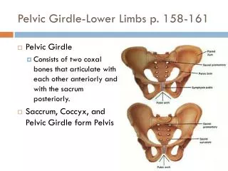
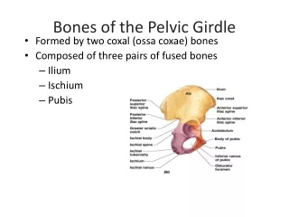
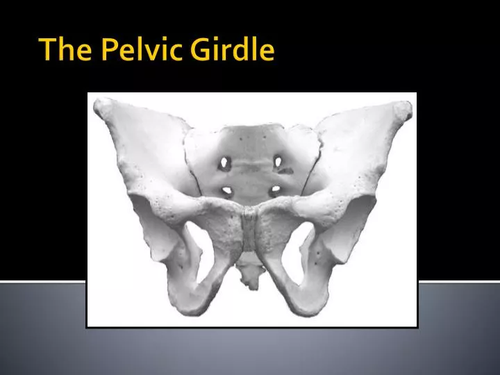
The Pelvic Girdle. Pelvic Girdle. Pelvis transmits weight of torso to legs Innominate “bone with no name” Also called the os coxae Firmly attached to axial skeleton at sacroiliac joint (to the sacrum) and to each other at pubic symphysis Anchors muscles for abdomen

E N D
Pelvic Girdle • Pelvis transmits weight of torso to legs • Innominate “bone with no name” • Alsocalled theoscoxae • Firmly attached to axial skeleton at sacroiliac joint (to the sacrum) and to each other at pubic symphysis • Anchors muscles for abdomen • Most reliable area for age and sex estimations from adults
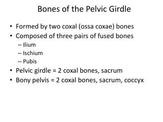
Ossification of Innominate • Each oscoxa is made up of 3 bones that meet at the acetabulum • Articular socket for femur – lateral • Where the 3 join is called the triradial strip (cartilage) • forms “Y-shape” • Within the socket is the lunate surface – where the femoral head actually articulates
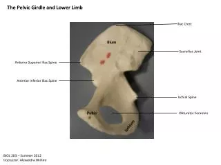
The 3 Parts of the Innominate • Ilium – superior – thin blade like upper portion • Ischium – inferior– blunt portion (what you sit on) • Pubis – ventral/anterior –where both sides meet in midline • Ischium and pubis join to enclose heart shaped opening called obturator foramen, which is closed with a membrane in life
Innominate • Ilium • Ischium • Pubis • Anterior superior iliac spine • Anterior inferior iliac spine
Innominate • Acetabulum • Iliac crest • Iliopubic ramus • Ischiopubic ramus • Pubic tubercle
Innominate • Pubic symphysis • Obturator foramen • Posterior superior iliac spine • Posterior inferior iliac spine • Auricular surface • Preauricular surface • Greater sciatic notch • Ischialspine • Lesser sciatic notch
Innominate • Iliac fossa
Pubic Symphysis • Irregular articular surface covered with hyaline cartilage and joined to opposing symphysis by fibro-cartilage • Ventral surface (external) • Dorsal surface • parturition pits
The Femur Proximal End (anterior) • Head • Neck • Intertrochanteric line
The Femur Proximal end (posterior) • Greater trochanter • Lesser trochanter • Intertrochanteric crest • Nutrient foramen • Linea aspera
The Femur Proximal end (medial) • Fovea capitis • Trochanteric fossa
The Femur Distal end (posterior) • Popliteal surface
The Femur Distal end (medial) Distal end (lateral) Lateral epicondyle Popliteal groove • Medial epicondyle
The Femur Distal end (inferior) • Patellar groove (surface) • Lateral condyle • Medial condyle • Intercondylar fossa
Ossification of Femur • 1 primary center • 4 secondary centers • Fuse during late teens to early 20s
The Patella • Apex • Medial articular facet • Lateral articular facet
Tibia Proximal (anterior) • Tibial tuberosity • Anterior crest
Tibia Proximal (posterior) • Tibial plateau • Popliteal (Soleal) line • Nutrient foramen
Tibia Proximal (superior) • Medial condyle • Lateral condyle • Intercondylar eminence
Tibia Lateral view • Interosseous crest • Fibular notch
Tibia Distal end (anterior) Distal end (posterior) Malleolar groove Distal end (inferior) • Medial malleolus • Talar facet
Fibula anterior • Head • Styloid process • Interosseous crest • Lateral malleolus
Fibula posterior • Fibular groove
Fibula medial • Nutrient foramen • Malleolar fossa
Ossification of Tibia & Fibula • 1 primary center • 2 secondary centers • Fuse during late teens to early 20s
Talocrural joint • Formed at articulation of tibia/fibula and talus (of the foot)
Radius Ulna Femur Tibia Fibula Humerus Lateral Medial