Download
1 / 8
210 likes | 1.17k Views
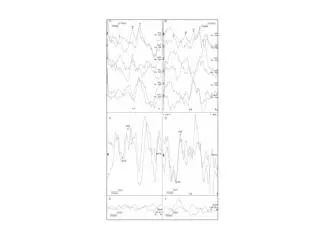
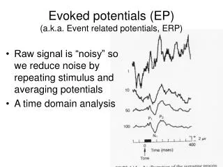
Evoked Potentials (EP). Sensory EP is a change in EEG resulting from stimulation of a sensory pathway Sensory EP is extracted from EEG using computer averaging techniques EEG is recorded during repetitive natural stimulation (eg. tap on skin or flash of light)

E N D
Evoked Potentials (EP) • Sensory EP is a change in EEG resulting from stimulation of a sensory pathway • Sensory EP is extracted from EEG using computer averaging techniques • EEG is recorded during repetitive natural stimulation (eg. tap on skin or flash of light) • Computers samples the EEG before & after stimulation & sample data are averaged.
Evoked Potentials (cont) • Sensory EP consist of multiple components related to various aspects of subcortical & cortical processing (scalp electrodes) • Clinically useful for assessing the function of sensory systems or evaluating demyelinating diseases (eg. M.S.) • Destruction of myelin causes conduction velocity to decrease which increase latencies
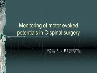
Somatosensory Evoked Potential • Repetative electrical stimulation of a peripheral nerve is used to elicit the SSEP which are recorded over the scalp & spine • Configuration & latency of the responses depend on the nerve that is stimulated
Clinical uses for EP • Detection of lesions in multiple sclerosis • functional status sometimes undetected with MRI • Detection of other CNS disorders • e.g. Spinocerebellar degeneration • Assessment & prognosis following CNS injury (trauma or hypoxia) • Intraoperative monitoring
Electromyography • The electrical activity within an accessible muscle can be recorded via insertion of a needle electrode into it. • Patterns of activity at rest and during contraction have been characterized under normal and abnormal conditions
Electromyography (EMG) • Activity at rest • Normal • no spontaneous electrical activity except at the end-plate region (neuromuscular junctions) • Abnormal • fibrillation potentials & positive sharp waves are associated with muscle fiber irritability & are typically found in denervated muscle or sometimes in myopathies (especially in inflammation) • fasciculation potentials - spntaneous activation of individual motor units (occasionally in normal muscle), characteristic of neuropathic disorders with primary involvement of anterior horn cells (e.g. ALS)
EMG • Activity during voluntary muscle contraction • Normal • a slight voluntary contraction of a muscle activates a small number of motor units • normal motor unit potentials have limits of duration, amplitude, configuration, & firing rates characteristic for muscle tested & # of motor units activated • Abnormal • in many myopathies- incidence small short duration polyphasic motor units & increased number of motor units activated for a given degree of voluntary activity • in neuropathies- loss of motor units # of units activated
Sensory nerve conduction studies • Stimulation of nerve to measure conduction velocity& amplitude of action potentials in sensory fibers when these fibers are stimulated at one point and response recorded at another point along course of the nerve • provide a means of confirming the presence & extent of peripheral nerve damage