Download
1 / 77
770 likes | 1.07k Views
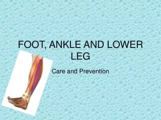
Chapter 19: The Ankle and Lower Leg. Functional Anatomy. Ankle is a stable hinge joint Medial and lateral displacement is prevented by the malleoli Ligament arrangement limits inversion and eversion at the subtalar joint Square shape of talus adds to stability of the ankle

E N D
Functional Anatomy • Ankle is a stable hinge joint • Medial and lateral displacement is prevented by the malleoli • Ligament arrangement limits inversion and eversion at the subtalar joint • Square shape of talus adds to stability of the ankle • Most stable during dorsiflexion, least stable in plantar flexion
Degrees of motion for the ankle range from 10 degrees of dorsiflexion to 50 degrees of plantar flexion • Normal gait requires 10 degrees of dorsiflexion and 20 degrees of plantar flexion with the knee fully extended • Normal ankle function is dependent on action of the rearfoot and subtalar joint
Preventing Injury in the Lower Leg and Ankle • Achilles Tendon Stretching • A tight heel cord may limit dorsiflexion and may predispose athlete to ankle injury • Should routinely stretch before and after practice • Stretching should be performed with knee extended and flexed 15-30 degrees • Strength Training • Static and dynamic joint stability is critical in preventing injury • While maintaining normal ROM, muscles and tendons surrounding joint must be kept strong
Neuromuscular Control Training • Can be enhanced by training in controlled activities • Uneven surfaces, BAPS boards, rocker boards, or Dynadiscs can also be utilized to challenge athlete
Footwear • Can be an important factor in reducing injury • Shoes should not be used in activities they were not made for • Preventive Taping and Orthoses • Tape can provide some prophylactic protection • However, improperly applied tape can disrupt normal biomechanical function and cause injury • Lace-up braces have even been found to be superior to taping relative to prevention
Assessing the Lower Leg and Ankle • History • Past history • Mechanism of injury • When does it hurt? • Type of, quality of, duration of pain? • Sounds or feelings? • How long were you disabled? • Swelling? • Previous treatments?
Observations • Postural deviations? • Genu valgum or varum? • Is there difficulty with walking? • Deformities, asymmetries or swelling? • Color and texture of skin, heat, redness? • Patient in obvious pain? • Is range of motion normal?
Fibular head and shaft Lateral malleolus Tibial plateau Tibial shaft Medial malleolus Dome of talus Calcaneus Peroneus longus Peroneus brevis Peroneus tertius Flexor digitorum longus Flexor hallucis Posterior tibialis Palpation: Bones and Soft Tissue
Anterior tibialis Extensor hallucis longus Extensor digitorum longus Gastrocnemius Soleus Achilles tendon Anterior/posterior talofibular ligament Calcaneofibular ligament Deltoid ligament Anterior tibiofibular ligament Posterior tibiofibular ligament Palpation: Soft Tissue (continued)
Special Test - Lower Leg • Lower Leg Alignment Tests • Malalignment can reveal causes of abnormal stresses applied to foot, ankle, lower leg, knees and hips • Anteriorly, a straight line can be drawn from ASIS, through patella and between 1st and 2nd toes • Laterally, a straight line can go from greater trochanter through center of patella and just behind the lateral malleolus • Posteriorly, a line can be drawn through the center of the lower leg, midline to the Achilles and calcaneus • Internal or external tibial torsion is also a common malalignment
Percussion and compression tests • Used when fracture is suspected • Percussion test is a blow to the tibia, fibula or heel to create vibratory force that resonates w/in fracture causing pain • Compression test involves compression of tibia and fibula either above or below site of concern • Thompson test • Squeeze calf muscle, while foot is extended off table to test the integrity of the Achilles tendon • Positive tests results in no movement in the foot • Homan’s test • Test for deep vein thrombophlebitis • With knee extended and foot off table, ankle is moved into dorsiflexion • Pain in calf is a positive sign and should be referred
Compression Test Percussion Test Homan’s Test Thompson Test
Ankle Stability Tests • Anterior drawer test • Used to determine damage to anterior talofibular ligament primarily and other lateral ligament secondarily • A positive test occurs when foot slides forward and/or makes a clunking sound as it reaches the end point • Talar tilt test • Performed to determine extent of inversion or eversion injuries • With foot at 90 degrees calcaneus is inverted and excessive motion indicates injury to calcaneofibular ligament and possibly the anterior and posterior talofibular ligaments • If the calcaneus is everted, the deltoid ligament is tested
Anterior Drawer Test Talar Tilt Test
Kleiger’s test • Used primarily to determine extent of damage to the deltoid ligament and may be used to evaluate distal ankle syndesmosis, anterior/posterior tibiofibular ligaments and the interosseus membrane • With lower leg stabilized, foot is rotated laterally to stress the deltoid • Medial Subtalar Glide Test • Performed to determine presence of excessive medial translation of the calcaneus on the talus • Talus is stabilized in subtalar neutral, while other hand glides the calcaneus, medially • A positive test presents with excessive movement, indicating injury to the lateral ligaments
Kleiger’s Test Medial Subtalar Glide Test
Functional Tests • While weight bearing the following should be performed • Walk on toes (plantar flexion) • Walk on heels (dorsiflexion) • Walk on lateral borders of feet (inversion) • Walk on medial borders of feet (eversion) • Hops on injured ankle • Passive, active and resistive movements should be manually applied to determine joint integrity and muscle function • If any of these are painful they should be avoided
Specific Injuries • Ankle Injuries: Sprains • Single most common injury in athletics caused by sudden inversion or eversion moments • Inversion Sprains • Most common and result in injury to the lateral ligaments • Anterior talofibular ligament is injured with inversion, plantar flexion and internal rotation • Occasionally the force is great enough for an avulsion fracture to occur w/ the lateral malleolus
Severity of sprains is graded (1-3) • With inversion sprains the foot is forcefully inverted or occurs when the foot comes into contact w/ uneven surfaces
Grade 1 Inversion Ankle Sprain • Etiology • Occurs with inversion plantar flexion and adduction • Causes stretching of the anterior talofibular ligament • Signs and Symptoms • Mild pain and disability; weight bearing is minimally impaired; point tenderness over ligaments and no laxity • Management • RICE for 1-2 days; limited weight bearing initially and then aggressive rehab • Tape may provide some additional support • Return to activity in 7-10 days
Grade 2 Inversion Ankle Sprain • Etiology • Moderate inversion force causing great deal of disability with many days of lost time • Signs and Symptoms • Feel or hear pop or snap; moderate pain w/ difficulty bearing weight; tenderness and edema • Positive talar tilt and anterior drawer tests • Possible tearing of the anterior talofibular and calcaneofibular ligaments • Management • RICE for at least first 72 hours; X-ray exam to rule out fx; crutches 5-10 days, progressing to weight bearing
Management (continued) • Will require protective immobilization but begin ROM exercises early to aid in maintenance of motion and proprioception • Taping will provide support during early stages of walking and running • Long term disability will include chronic instability with injury recurrence potentially leading to joint degeneration • Must continue to engage in rehab to prevent against re-injury
Grade 3 Inversion Ankle Sprain • Etiology • Relatively uncommon but is extremely disabling • Caused by significant force (inversion) resulting in spontaneous subluxation and reduction • Causes damage to the anterior/posterior talofibular and calcaneofibular ligaments as well as the capsule • Signs and Symptoms • Severe pain, swelling, hemarthrosis, discoloration • Unable to bear weight • Positive talar tilt and anterior drawer
Management • RICE, X-ray (physician may apply dorsiflexion splint for 3-6 weeks) • Crutches are provided after cast removal • Isometrics in cast; ROM, PRE and balance exercise once out • Surgery may be warranted to stabilize ankle due to increased laxity and instability
Eversion Ankle Sprains -(Represent 5-10% of all ankle sprains) • Etiology • Bony protection and ligament strength decreases likelihood of injury • Eversion force results in damage to deltoid ligament and possibly fx of the fibula • Deltoid can also be impinged and contused with inversion sprains
Etiology (continued) • Due to severity of injury, it may take longer to heal • Foot that is pronated, hypermobile or has a depressed medial longitudinal arch is more predisposed to eversion sprains • Signs and Symptoms • Pain may be severe; unable to bear weight; and pain with abduction and adduction but not direct pressure on bottom of foot • Management • RICE; X-ray to rule out fx; no weight bearing initially; posterior splint tape; NSAID’s • Follows the same course of treatment as inversion sprains • Grade 2 or higher will present with considerable instability and may cause weakness in medial longitudinal arch resulting in excessive pronation or fallen arch
Syndesmotic Sprain • Etiology • Injury to the distal tibiofemoral joint (anterior/posterior tibiofibular ligament) • Torn w/ increased external rotation or dorsiflexion • Injured in conjunction w/ medial and lateral ligaments • Signs and Symptoms • Severe pain, loss of function; passive external rotation and dorsiflexion cause pain • Pain is usually anterolaterally located • Management • Difficult to treat and may requires months of treatment • Same course of treatment as other sprains, however, immobilization and total rehab may be longer
Ankle Fractures/Dislocations • Etiology • Number of mechanisms • Signs and Symptoms • Swelling and pain may be extreme with possible deformity • Management • RICE to control hemorrhaging and swelling • Once swelling is reduced, a walking cast or brace may be applied, w/ immobilization lasting 6-8 weeks
Osteochondritis Dissecans • Etiology • Occur in superior medial articular surface of the talar dome • One or several fragments of articular cartilage, w/ underlying subchondral bone partially or completely detached and moving within the joint space • Mechanism may be single trauma or repeated traumas • Signs and Symptoms • May be a complaint of pain and effusion with signs of atrophy • May also be catching, locking, or giving way
Osteochondritis Dissecans • Management • Diagnosis through X-ray or MRI • Incomplete and non-displaced injuries can be immobilized with early motion and delayed weight bearing • If fragments are displaced, surgery is necessary • Surgery will minimize risk of nonunion
Acute Achilles Strain • Etiology • Common in sports and often occurs with sprains or excessive dorsiflexion • Sign and Symptoms • Pain may be mild to severe • Most severe injury is partial/complete avulsion or rupturing of the Achilles • Management • Pressure and RICE should be applied • After hemorrhaging has subsided an elastic wrap should continue to be applied • Conservative treatment should be used as Achilles problems generally become chronic • A heel lift should be used and stretching and strengthening should begin soon
Achilles Tendinitis • Etiology • Inflammatory condition involving tendon, sheath or paratenon • Tendon is overloaded due to extensive stress • Presents with gradual onset and worsens with continued use • Decreased flexibility exacerbates condition • Signs and Symptoms • Generalized pain and stiffness, localized proximal to calcaneal insertion • Warm and painful with palpation, also presents with thickening • May limit strength • May progress to morning stiffness
Crepitus with active plantar flexion and passive dorsiflexion • Chronic inflammation may lead to thickening • Management • Resistant to quick resolution due to slow healing nature of tendon • Must reduce stress on tendon, address structural faults (orthotics, mechanics, flexibility) • Use anti-inflammatory modalities and medications • Cross friction massage may be helpful in breaking down adhesions • Strengthening must progress slowly in order to not aggravate the tendon
Achilles Tendon Rupture • Etiology • Occurs w/ sudden stop and go; forceful plantar flexion w/ knee moving into full extension • Commonly seen in athletes > 30 years old • Generally has history of chronic inflammation • Signs and Symptoms • Sudden snap (kick in the leg) w/ immediate pain which rapidly subsides • Point tenderness, swelling, discoloration; decreased ROM • Obvious indentation and positive Thompson test • Occurs 2-6 cm proximal the calcaneal insertion
Achilles Tendon Rupture (continued) • Management • Usual management involves surgical repair for serious injuries (return of 75-80% of function) • Non-operative treatment consists of RICE, NSAID’s, analgesics, and a non-weight bearing cast for 6 weeks, followed up by a walking cast for 2 weeks (75-90% return to normal function) • Rehabilitation lasts about 6 months and consists of ROM, PRE and wearing a 2cm heel lift in both shoes
Peroneal Tendon Subluxation/Dislocation • Etiology • Occurs in sports with dynamic forces being applied to the ankle • May also be caused by dramatic blow to posterior lateral malleolus, or moderate/severe inversion ankle sprain resulting in tearing of peroneal retinaculum • Signs and Symptoms • Complain of snapping in and out of groove with activity • Eversion against manual resistance replicates subluxation • Recurrent pain, snapping and instability • Present with ecchymosis, edema, tenderness, and crepitus over the tendon
Peroneal Subluxation (continued) • Management • Conservative approach should be used first, including compression with felt horseshoe • Reinforce compression pad with rigid plastic or plaster until acute signs have subsided • RICE, NSAID’s and analgesics • Conservative treatment time 5-6 weeks followed by gradual rehab program • Surgery if conservative plan fails