Download

1 / 62
670 likes | 994 Views
Novak 2003. Gestational trophoblastic disease. Hydatidiform Mole Persistent Gestational Trophoblastic Tumor Chemotherapy. Hydatidiform mole. Epidemiology Complete versus partial mole Clinical picture Natural history Diagnosis

E N D
Novak 2003 Gestational trophoblastic disease
Hydatidiform Mole • Persistent Gestational Trophoblastic Tumor • Chemotherapy
Epidemiology • Complete versus partial mole • Clinical picture • Natural history • Diagnosis • Treatment • Follow up
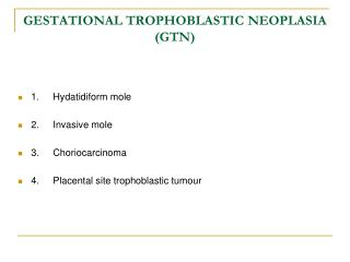
GTD is among the rare tumors that can be cured even if metastasized Types: • Complete mole • Partial mole • Placental site mole • Choriocarcinoma Persistent GTT: Most commonly follow molar pregnancy May also follow: abortion, ectopic or term pregnancy introduction
% varies in different sites: Japan = 2 : 1000 pregnancies USA = 0.6 – 1.1 : 1000 pregnancies In pathological studies: Complete mole 1 : 945 Partial mole 1 : 695 epidemiology
Risk factors in complete mole: 1 – nutritional: ↓ carotene ↓ vit A 2 – Age: > 35 years = X 2 > 40 years = X 7.5 Risk factors in partial mole: 1 - OCCP 2 - H/O irregular menstruation
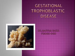
Complete mole Pathology: • No fetal or embryonic tissue • Villi show: Diffuse hydropic swelling Diffuse trophoblastic hyperplasia Chromosome: 90% 46XX 10% 46XY Complete versus partial mole
Chromosomes are entirely paternal Mitochondria DNA is maternal in origin 1 - Absent or inactivated ovum nucleus + 1 haploid sperm endoredublication homozygous mole 2– Absent or inactivated ovum nucleus + 2 haploid sperms heterozygous mole
Partial mole Villi vary in size and show: • Focal hydropic swelling • Focal trophoblastic hyperplasia • Focal cavitation • Stromaltrophoblastic inclusion • Scalloping Fetal or embryonic tissues
Chromosomes: Absent or inactivated ovum nucleus + 3 haploid sperms triploid in 90% = 69XXX, 69XXY, 69XYY The fetus shows triploidy stigmata: • GR • Multiple congenital anomalies as: Syndactyly - Hydrocephalus
Complete Partial Fetus absent present Karyotype 46XX(90%) 69XXX 46XY (90%) Hydropic swelling diffuse focal Trophoblastic diffuse focal hyperpleasia Scalloping no present Stromal inclusions no present
Complete Partial past now Vaginal bleeding 97% 84% 74% Anemia 50% 5% Excessive uterine size 50% 28% 4% Preeclampsia 50% 1.5% Hyperemesis 27% 8% Hyperthyroidism 7% 0% Trophoblastic embolism 2% 0% Theca lutein cysts 50% HCG > 100,000mIU/mL 6% Clinical picture
Excessive uterine size: = ↑ trophoblastic tissue ↑ hCG ↑ preeclampsia ↑ hyperthyroidism ↑ hyperemesisgravidarum ↑ trophoblasticembolization ↑ theca lutein cyst size
Preeclampsia: Early preeclampsia = hydatidiform mole Hyperthyroidism: Due to ↑ free T3, T4 C/P: • tachycardia • warm skin • tremor
Thyroid storms: Give β–blockers before anesthesia to avoid thyroid storms C/P: ↑ pulse, ↑ temp, ↑ COP + delirium + convulsions may HF
Trophoblasticembolization: C/P: • dyspnea • cough • tachypnea • ↑ P • chest pain • asymptomatic
Chest examination diffuse rales Chest X ray bilateral infiltrates Causes of respiratory distress: • Trophoblastionembolization • Complications of: • preeclampsia • thyroid storm • excessive fluid intake
Theca lutein ovarian cysts • Due to ovarian overstimulation by ↑ hCG • May not be felt with oversized uterus • May pressure symptoms treated by decompression by laparoscopic or U/Sguided aspiration • If ruptured or torsion occur acute pain laparoscope
Complete mole • Invasive = 15% • Metastatic = 4% Risk factors: • hCG > 100,000 mIU/mL • Excessive uterine size • Theca lutein cysts = 6 cm Natural history
Low risk = 60% 3.4% persistent mole 0.6% metastatic High risk = 40% 31% persistent mole 9% metastatic Age: > 40 years = 37% > 50 years = 56%
Complete mole U/S vesicular pattern Partial mole U/S focal cystic spaces in placenta + ↑ transverse diameter of GS Both together 90% +ve predictive value diagnosis
I – Hystrectomy + aspiration of CL cyst + follow up as usual 2 - Suction evacuation Preferred ttt for hydatidiform mole Give oxytocine before anesthesia Use 12 canula If > 14 weeks support the fundus + do fundal massage treatment
Dilatation ↑ bleeding Suction evacuation ↓ bleeding If RH –ve give Anti RH Ig 3 - Prophylactic chemotherapy ↓ invasive mole to 4% after 1stcourse ↓ “””””””””””””””””” 0% after 2nd course Controversial : Why to expose all patients to chemotherapy while only 20% will need it?
Useful if follow up is: Unreliable Unavailable Study: Prophylactic chemotherapy in high risk patients ↓ persistent mole from 47% to 14%
1 - HCG Average time needed to return to normal values = 9 weeks Measure hCG/week 3 consecutive normal results /month 6 consecutive normal R 2 - Contraception: OCCP or barrier methods IUD is C/I perforation Follow up
Nonmetastatic disease • Placental-site TT • Metastatic D • Staging • Prognostic scoring systems • Diagnostic evaluation • Management
Invasive mole = 15% after evacuation C/P: • Irregular vaginal bleeding • Uterine subinvolution • Theca lutein cysts • ↑hCG • Perforation of myometrium internal Hg • Perforation of uterine vessels vaginal Hg • Infection acute pain purulent discharge Nonmetastatic disease
Histology: • After molar pregnancy hydatidiform mole or choriocarcinoma • After nonmolar pregnancy choriocarcinoma = sheets of anaplastic cytotrophoblast and syncytiotrophoblast + no villi
Uncommon Variant of choriocarcinoma Consists of intermediate trophoblast Produce small amounts of hCG & hPL Tends to be confined to the uterus Metastasize late Resistant to chemotherapy Placental-site tt
= 4% after molar pregnancy More often after nonmolar pregnancy Usually associated with choriocarcinoma Highly vascular spontaneous bleeding Early vascular spreading Sites: Pulmonary 80% Hepatic 10% Vaginal 30% Brain 10% Pelvic 20% Metastatic disease
1 – Pulmonary metastases: Symptoms: dyspnea cough hemoptysis chest pain asymptomatic May be acute of chronic
Chest X ray: • Snowstorm pattern • Discrete rounded densities • Pleural effusion • Pulmonary artery embolism May be misdiagnosed as 1ry pulmonary disease and only recognized as GTD after thoracotomy
Pulmonary embolism may pulmonary HTN Early RF + intubation = bad prognosis 2 – Vaginal metastasis highly vascular biopsy may excessive bleeding Symptoms: Vaginal bleeding Purulent discharge Site: fornices/suburethral
3 – Hepatic metastasis Usually in advanced cases Symptoms: Epigastric or upper RT ¼ pain due to stretching subcapsular hematoma Rupture internal Hg 4 – Brain metastasis Usually in advanced cases Spontaneous bleeding acute focal neurological defects
Stage I confined to uterus Stage II confined to genital structures Stage III pulmonary metastasis Stage IV other metastasis At any stage: A= no risk factors B= 1 risk factor C= 2 risk factors staging
0 1 2 4 Age ≤39 >39 Pregnancy mole abortion term Duration <4m 4-6 7-12 >12 hCG <1000 <10000 <100000 > Largest size <3cm 3-5 >5 Site of met 0 kidney/spleen GIT/liver brain Number <3 1-3 4-8 >8 ABO group 0 A/O B/AB Chemotherapy 1 ≥2 Prognostic scoring systems
H/O • Examination • hCG • Liver function tests • Kidney function tests • Thyroid function tests • WBCs • Platelet count Diagnostic evaluation
Chest X-ray -- CT Abd & pelvis U/S -- CT Brain MRI -- CT If no pulmonary or vaginal metastasis metastasis are rare Chest CT for micrometastasis Liver CT for abnormal LFTs Brain CT for asymptomatic lesions imaging
If brain CT is normalmeasure CSF hCG If serum hCG/CSF hCG = < 60% then there is brain metastasis Pelvic U/S for: • Extent of uterine lesion • Localization of resistant lesions • Identifying patients who will benefit from hystrectomy
Stage I • Stage II & III • Stage IV management
If the patient does not wish to preserve fertility Hystrectomy + Chemotherapy to: • ↓ dissemination of GTD • Treat dissemination of GTD • Treat occult metastasis • ↓ bleeding • ↓ sepsis Stage I
If the patient wish to preserve fertility: Low risk Single agent High risk Combined chemotherapy Resistant Local uterine resection after localization of resistant sites by U/S, MRI, or arteriography
Placental site GTD: - Only curative ttt for nonmetastatic cases is hystrectomy - Resistant to chemotherapy few metastatic cases reported complete remission after chemotherapy
Pulmonary metastasis Low risk single agent 82% CR High risk combined chemotherapy Resistant thoracotomy after localization of and exclusion of other resistant sites Stage ii & iii
Vaginal metastasis Low risk single agent 84% CR High risk combined chemotherapy Resistant wide local excision Vaginal bleeding: • Packing of the vagina • Wide local excision • Embolization of hypogastric arteries
Hystrectomy - In metastatic disease - to control Hg - to control sepsis - In extensive uterine disease - to ↓ GTT burden - to ↓ chemotherapy courses
Follow up of stage I, II, III: hCG/week 3 consecutive normal results hCG/month 12 consecutive normal results + effective contraception