Download
1 / 1
10 likes | 104 Views
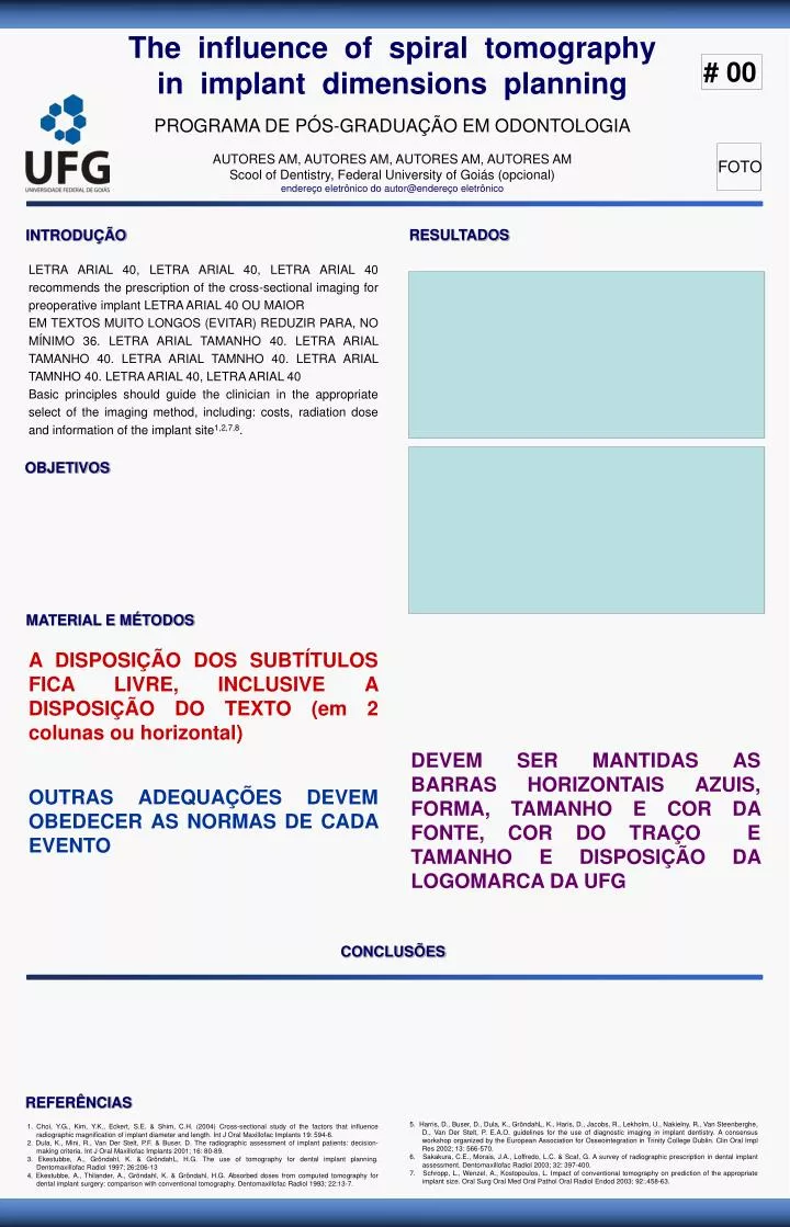
The influence of spiral tomography in implant dimensions planning. # 00. PROGRAMA DE PÓS-GRADUAÇÃO EM ODONTOLOGIA. AUTORES AM, AUTORES AM, AUTORES AM, AUTORES AM Scool of Dentistry, Federal University of Goiás (opcional) endereço eletrônico do autor@endereço eletrônico. FOTO.

E N D
The influence of spiral tomography in implant dimensions planning # 00 PROGRAMA DE PÓS-GRADUAÇÃO EM ODONTOLOGIA AUTORES AM, AUTORES AM, AUTORES AM, AUTORES AM Scool of Dentistry, Federal University of Goiás (opcional) endereço eletrônico do autor@endereço eletrônico FOTO RESULTADOS INTRODUÇÃO LETRA ARIAL 40, LETRA ARIAL 40, LETRA ARIAL 40 recommends the prescription of the cross-sectional imaging for preoperative implant LETRA ARIAL 40 OU MAIOR EM TEXTOS MUITO LONGOS (EVITAR) REDUZIR PARA, NO MÍNIMO 36. LETRA ARIAL TAMANHO 40. LETRA ARIAL TAMANHO 40. LETRA ARIAL TAMNHO 40. LETRA ARIAL TAMNHO 40. LETRA ARIAL 40, LETRA ARIAL 40 Basic principles should guide the clinician in the appropriate select of the imaging method, including: costs, radiation dose and information of the implant site1,2,7,8. OBJETIVOS MATERIAL E MÉTODOS A DISPOSIÇÃO DOS SUBTÍTULOS FICA LIVRE, INCLUSIVE A DISPOSIÇÃO DO TEXTO (em 2 colunas ou horizontal) DEVEM SER MANTIDAS AS BARRAS HORIZONTAIS AZUIS, FORMA, TAMANHO E COR DA FONTE, COR DO TRAÇO E TAMANHO E DISPOSIÇÃO DA LOGOMARCA DA UFG OUTRAS ADEQUAÇÕES DEVEM OBEDECER AS NORMAS DE CADA EVENTO CONCLUSÕES REFERÊNCIAS 5. Harris, D., Buser, D., Dula, K., GröndahL, K., Haris, D., Jacobs, R., Lekholm, U., Nakielny, R., Van Steenberghe, D., Van Der Stelt, P. E.A.O. guidelines for the use of diagnostic imaging in implant dentistry. A consensus workshop organized by the European Association for Osseointegration in Trinity College Dublin. Clin Oral Impl Res 2002; 13: 566-570. 6. Sakakura, C.E., Morais, J.A., Loffredo, L.C. & Scaf, G. A survey of radiographic prescription in dental implant assessment. Dentomaxillofac Radiol 2003; 32: 397-400. 7. Schropp, L., Wenzel, A., Kostopoulos, L. Impact of conventional tomography on prediction of the appropriate implant size. Oral Surg Oral Med Oral Pathol Oral Radiol Endod 2003; 92:.458-63. 1. Choi, Y.G., Kim, Y.K., Eckert, S.E. & Shim, C.H. (2004) Cross-sectional study of the factors that influence radiographic magnification of implant diameter and length. Int J Oral Maxillofac Implants 19: 594-6. 2. Dula, K., Mini, R., Van Der Stelt, P.F. & Buser, D. The radiographic assessment of implant patients: decision-making criteria. Int J Oral Maxillofac Implants 2001; 16: 80-89. 3. Ekestubbe, A., Gröndahl, K. & GröndahL, H.G. The use of tomography for dental implant planning. Dentomaxillofac Radiol 1997; 26:206-13 4. Ekestubbe, A., Thilander, A., Gröndahl, K. & Gröndahl, H.G. Absorbed doses from computed tomography for dental implant surgery: comparison with conventional tomography. Dentomaxillofac Radiol 1993; 22:13-7.