Download
1 / 1
10 likes | 90 Views
COUPLING A TUNABLE OPO LASER TO AN FT- ICR MASS SPECTROMETER FOR INFRARED PHOTODISSOCIATION STUDIES J. Szczepanski, E. Cagmat, W. Mino, W. Pearson, D. Powell, J.R. Eyler, N. Polfer Department of Chemistry, University of Florida, Gainesville, FL 32611-7200, USA. Introduction. Results.

E N D
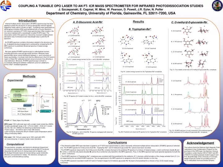
COUPLING A TUNABLE OPO LASER TO AN FT- ICR MASS SPECTROMETERFOR INFRARED PHOTODISSOCIATION STUDIES J. Szczepanski, E. Cagmat, W. Mino, W. Pearson, D. Powell, J.R. Eyler, N. Polfer Department of Chemistry, University of Florida, Gainesville, FL 32611-7200, USA Introduction Results A. D-Glucuronic Acid-Rb+ C. O-methyl-β-D-glucoside-Rb+ Infrared multiple-photon dissociation (IR-MPD) spectroscopy has been applied to probe the structures of various biologically relevant ligands tagged by metal cations (shown at right). Complexes were generated via electrospray ionization (ESI) and transferred into a Fourier transform – ion cyclotron resonance (FT-ICR) mass spectrometer. After isolation, the ions were irradiated with a wavelength-tunable IR OPO laser. The dissociation yield of the parent ion induced by resonant multiple-photon absorption was monitored at each wavelength step to yield the IR spectrum. An IR-MPD spectrum contains information related to the structure of an ion. A detailed structural assignment is achieved by comparing the IR-MPD spectrum to predicted vibrational spectra of lowest energy conformers. We have applied IR-MPD spectroscopy to carbohydrate isomers, including glucuronic acid and glucoside anomers. Whereas mass spectrometry alone cannot distinguish between a- and b-anomers of these saccharides, it is shown here that the differences in the O-H stretch region of these Rb+-adducted isomers are pronounced, thus allowing a clear distinction. Moreover, IR-MPD spectroscopy is shown to successfully distinguish between zwitterionic and non-zwitterionic forms of Ba2+-chelated tryptophan. B. Tryptophan-Ba2+ A. (0.0 kcal/mol) a-3 (2.82 kcal/mol) b-3 (2.83 kcal/mol) B. (1.79 kcal/mol) b-2 (1.87 kcal/mol) a-2 (1.52 kcal/mol) Z-2 (0.4 kcal/mol) Z-3 (6.7 kcal/mol) C. (5.19 kcal/mol) b-1 (0.00 kcal/mol) a-1 (0.00 kcal/mol) Fig. A1. Lowest energy isomers for the a- and b-GlcA•Rb+ complexes D. (6.49 kcal/mol) a-3 (2.82 kcal/mol) b-3 (2.83 kcal/mol) Z-1 (0.0 kcal/mol) C1 (8.0 kcal/mol) Fig. C1. Four stable conformers of O-methyl-β-D-glucoside-Rb+ complexes Integral Intensities (km/mol) Fig. B1. Lowest energy isomers for the Tryptophan-Ba2+complexes b-2 (1.87 kcal/mol) a-2 (1.52 kcal/mol) Methods 2 1 • 1. O-H • 2. (N-H)ring • 3. (N-H)as • 4. (N-H)s Experimental b-1 (0.0 kcal/mol) A. (0.0 kcal/mol) a-1 (0.0 kcal/mol) C1 (8.0 kcal/mol) 4 3 Wavemeter Fragment Yield MPD (exp) MPD (exp) ICR Cell Z-3 (6.7 kcal/mol) Wavenumbers (cm-1) Wavenumbers (cm-1) B. (1.79 kcal/mol) Fig. A2. IR spectra (O-H stretch modes) for the a- and b-GlcA•Rb+ complexes ESI Integral Intensities (km/mol) 4.7 T Magnet b-1 a-1 Z-2 (0.4 kcal/mol) MPD Power Meter D CO2 Laser Uniblitz Beam Reflecting Shutter MPD band Assignment 1 3 • 1. (N-H)ring • 2. (N-H)as • 3. (N-H)s+(N-H)out-ph • 4. (N-H)s+(N-H)in-ph IR Idler Beam A (a + b) B a C b D (a + b) C. (5.19 kcal/mol) Z-1 (0.0 kcal/mol) 2 OPO Laser NIR Signal Beam 4 A IR, 2nd Step Excitation C Intensity (a. u.) B FT-ICR: 4.7 Tesla (Apex II by Bruker) OPO Laser: CW multimode laser with a single mode locked (by LINOS). Tunable wavelength range, 2.32 - 4.76 mm (4300 – 2100 cm-1) covers the OH, NH, CH, OHN(or O) stretching fundamental modes. Power output, ~ 60 mW for each of two idler beams. Laser tuning by varying the lithium niobate crystal temperature and/or tilting a intracavity etalon. CO2 Laser: CW, 10.6 μm output up to 15W, used with OPO as a second pump, in cases of weak mode intensities. MPD (exp) Fragment Yield D. (6.49 kcal/mol) Wavenumbers (cm-1) Wavenumbers (cm-1) Fig. C2. IR-MPD spectrum (in red) compared to the predicted spectra of four conformers of O-methyl-β-D-glucoside-Rb+ complexes Fig. B2. Predicted and observed IR spectra for the Tryptophan-Ba2+complexes Fig. A3. Predicted a- and b-GlcA•Rb+ IR spectra overlapped with observed IR-MPD spectrum Conclusions Acknowledgement Computational • The infrared tunable OPO laser has been coupled to an FT-ICR mass spectrometer to collect resonantly enhanced multiple-photon dissociation (IR-MPD) spectra of selected ions. The IR-MPD spectra for D-Glucuronic Acid-Rb+, Tryptophan-Ba2+ and O-methyl-β-D-glucoside-Rb+cations have been recorded. • The IR-MPD spectrum observed for the D-Glucuronic Acid-Rb+ complex fits best to the predicted IR spectra of a mixture of lowest energy a- and b-conformers (bands A and D), to the a-conformer only (band shoulder B) and to the b-conformer only (band C in the MPD spectrum). Thus, in the MPD spectrum we found spectral signatures for both the a- and b- conformers. • The IR-MPD spectrum of Tryptophan-Ba2+ complexes was consistent with that predicted for the zwitterion (Z-1) structure, while formation of the charge solvated form (C-1) is not supported by the recorded IR-MPD spectrum. The strongest band in the MPD spectrum is assigned to the N-H stretch vibration on the ring. •The IR-MPD spectrum recorded in both O-H and C-H stretch ranges for O-methyl-b-glucoside-Rb+ fits best to that predicted for a mixture of four (A-D) lowest energy conformers. The geometries, energies, and harmonic vibrational frequencies of complexes studied were calculated using the B3LYP functional (Gaussian 03) with the 6-31+G(d,p) basis set for ligands, and the Stuttgart-Dresden basis set incorporating a quasi-relativisitic effective core potential for metals. The electronic energies are zero point corrected without scaling. The authors thank the National High Magnetic Field Laboratory's User Collaboration Program for the purchase of the OPO laser, the National Science Foundation (under Grants CHE-0718007 and INT-0730072) for partial graduate student support and Prof. Brad Bendiak of the University of Colorado Health Sciences Center for providing some of the O-methylated monosaccharide samples.