Download
1 / 37
460 likes | 668 Views
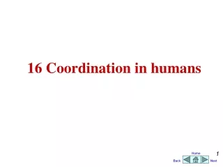
Coordination and Response Nervous control in Humans. * Describe the structure of the nervous system **Distinguish between voluntary and involuntary actions. Homework: Revise Homeostasis and Excretion for test on 3/5/11. Blinking. We think about this action/ We don’t think about this action.

E N D
Coordination and ResponseNervous control in Humans *Describe the structure of the nervous system **Distinguish between voluntary and involuntary actions Homework: Revise Homeostasis and Excretion for test on 3/5/11
Blinking We think about this action/ We don’t think about this action
Coughing We think about this action/ We don’t think about this action
Kicking We think about this action/ We don’t think about this action
Pupils changing size We think about this action/ We don’t think about this action
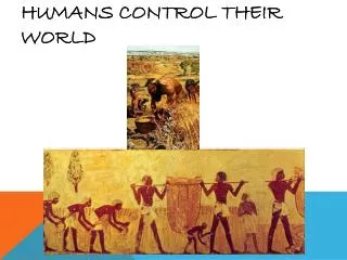
All living organisms are sensitive to changes in their environment. The changes they detect are called stimuli. E.g. ? The cells that detect these changes are called receptors. E.g.? The responses are brought about by muscles and glands because of a stimuli and they are called effectors. Which system is responsible for the detection? Which system is responsible for the coordination of responses ? sensory nervous
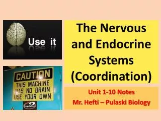
Human Nervous System Brain Protected by? i Cranial Nerves connected in pairs to the brain CNS Spinal Cord Protected by The nerves are made up of specialised cells called neurons Cranial nerves link the brain to all organs in the head and some organs of the abdomen and thorax Spinal nerves link brain to legs ,arms , thorax and abdomen PNS Spinal nerves connected in pairs to the spine .
Voluntary Response . Effector • Voluntary responses are under the control of our brains. For example; Response Motor Neuron Stimulus Coordinator Receptor Transmits electrical impulses @ 1-120m/s Sensory neuron Spinal Cord
Some facts • Bundles of neurons are called nerves. • The neurons joining the sensory and the motor neurons are called relay neurons • Effectors are muscles or glands.
Putting it all together! coordinator Receptor --> sensory neurone → CNS(relay neuron) → motor neurone → effector. Write one more examples of voluntary action. In the last example who was the receptor and who was the effector?
Involuntary Response or Reflex Action • What is an involuntary response? • What do you think will happens to the man’s hand? • http://www.bbc.co.uk/schools/gcsebitesize/science/ocr_gateway/ourselves/3_keeping_in_touch6.shtml
Involuntary Response or Reflex Action • An involuntary response bypasses the brain to give a fast response to a stimulus. • This helps protect the body from harm.
Involuntary Response Effector For example; Motor Neurone Stimulus Receptor Transmits impulses Sensory neurone Spinal Cord
Give one more examples of reflex action. Draw one of your examples from both voluntary and involuntary actions as a flow diagram.
Sense Organs and Reflex Arcs Objectives: *Define sense organs ** Describe their functioning in a reflex action Starter: Complete the flow diagram 1.Receptor - - - - - - - - - - - - - - - - - - - 2. What are five senses in your body?
Brentwood Gazette Weekly Rainbow Control of a possible football situation RISU are trailing 1-0 to ISU in the final of the Rainbow Cup. All of a sudden (student A) pulls up with a suspected torn hamstring. (Student B) spots this and immediately sends a message to Mr Colley what happens. Mr Colley makes a decision. He decides to replace (student A) with (Student C). Student C went on to score a hat trick. Final Score RISU 3 ISU 4 In terms of response: 1.What kind of response is this? 2. What was the stimulus? 3. What was the effector? 4. What was the response?
Nervous system Parts of our body we use to sense things are called SENSE ORGANS – eye, ear, mouth, skin, nose. Each SENSE ORGAN has special cells called RECEPTOR CELLS. Each receptor cell is sensitive to different things. SENSE ORGANS are a group of receptor cells that respond to a particular stimuli
Senses • Receptors in eye sensitive to light • Receptors in ear and sensitive to sound • Receptors on tongue and sensitive to chemicals • Receptors in nose and sensitive to chemicals • Receptors in the skin and sensitive to touch, pressure, pain, temperature
Reflex actions A receptor detects a stimulus. The receptor sends an electrical impulse along a sensory neuron • The tap on the knee in the knee jerk test is a stimulus. • Its detected by receptors in the thigh muscle connected to your knees. • The receptor sends signals to your spinal cord. • The spinal cord sends nerve impulses to your leg muscles. • The leg muscles respond by contracting which pulls your lower leg upwards. In a reflex action: These impulses are sent to the CNS. The CNS sends an electrical impulse along a motor neuron to an effector. The effector responds to the stimulus. A reflex action is a fast, automatic response to a stimulus.
Reflex Arc • Stimulus picked up by Receptor cells in finger (skin) • Impulse passed on to sensory neuron • Sensory neuron passes impulse to spinal cord • Spinal cord sorts out response and send message to motor neuron • Motor neuron sends impulses to finger muscles to pull away stimulus Relay neuron effector
3 Spinal cord or brain sorts out message Impulse carried along nerve cell (motor neurone) to effector organ 4 Effector organ brings about a response 5 Stimulus (change) picked up by receptor 1 Impulse carried along nerve cell (Sensory neurone) to spinal cord 2 Arrange them in right order
Neurones Objectives: *Describe and distinguish between three types of neuron **Describe the functioning of these neurons in a reflex action Starter: How many neurons are there? Name them.
Relay neurons short pass on impulses from sensory to motor neuron located inside the CNS Neurons • It carries information from the nervous system as electrical impulses. • These cell that carry this information are called nerve cells or neurones. Nerve ending in a sense organ nucleus cytoplasm • This is a sensory neuron. • It carries information from the receptor to the CNS. (Insulation) Neurone ending in an effector(muscleor gland) in CNS • This is a motor neuron. • It carries information from the CNS to the effector.
The Structure of the Neuron • Cell bodycontains nucleus cytoplasm and nerve fibres • Fibres carrying impulses away from cell body are Axons • Fibres carrying impulses towards cell body are Dendrons with smaller Dendritesextending from cell body • The Myelin Sheathis a thick insulating material (fat) that encloses the axon. It enables fast conduction of impulses up to 100m/s • to fast transmission)
The neurones do not touch each other. The gap between them is called synapse • Impulse arrives at a synapse in a particular direction • Chemical molecules released by the sensory neuron diffuses across and fits on to the receptor molecules on the membrane of the motor neuron • Nerve impulse passes from sensory neuron to motor neuron • The chemical is absorbed back in the sensory neuron • Since chemicals are produced on one side impulses travel in only one direction • Many drugs produce their effects by acting at synapses Some facts: Synapses are 20 nm wide and slows down the speed of your impulse by 15m/s
Reflex actions A receptor detects a stimulus. The receptor sends an electrical impulse along a sensory neurone • The tap on the knee in the knee jerk test is a stimulus. • Its detected by receptors in the thigh muscle connected to your knees. • The receptor sends signals to your spinal cord. • The spinal cord sends nerve impulses to your leg muscles. • The leg muscles respond by contracting which pulls your lower leg upwards. In a reflex action: These impulses are sent to the CNS. The CNS sends an electrical impulse along a motor neurone to an effector. The effector responds to the stimulus. A reflex action is a fast, automatic response to a stimulus.
Reflexes and Reaction time Objectives: *Explain the importance of reflex action in the body ** Calculate reaction time *** Represent data in different ways Starter: Reflex actions are a…….., f… and usually p……… fast protective automatic
Reaction time The time between a stimulus and a response Eg. If someone is driving and they see a cow in the middle of the road. Their reaction time is the time taken to see the cow (stimulus)and to press the brake (response).
Reaction time: Practical http://www.bbc.co.uk/science/humanbody/sleep/sheep/
How fast are your reflexes?? • How can we measure our reflexes? Practical experiment….Using a ruler 1.Using a ruler you have to see how quickly you can catch it. 2. Each person will try three times and record their results in a table 3. We will then collect the class data and find out who has the fastest reflexes!!!
Results NOTE : To work out the average you add all the result up, then divide by three…….EASY!!
How else can we display our results? Remember Title, label axis, 0 Who is the fastest - Class results Graph


![Nervous System [humans]](https://cdn1.slideserve.com/2761942/nervous-system-humans-dt.jpg)