Download
1 / 46
500 likes | 845 Views
Part 2. DNA Lecture . Some of the following slides and text are taken from the DNA Topology lecture from Doug Brutlag’s January 7, 2000 Biochemistry 201 Advanced Molecular Biology Course at Stanford University. DNA Topology. What Is Supercoiling & Why Should I Care?.

E N D
Part 2 DNA Lecture
Some of the following slides and text are taken from the DNA Topology lecture from Doug Brutlag’s January 7, 2000 Biochemistry 201 Advanced Molecular Biology Course at Stanford University DNA Topology
What Is Supercoiling & Why Should I Care? • DNA forms supercoils in vivo • Important during replication and transcription • Topology only defined for a continuous strand - no strand breakage • Numerical expression for degree of supercoiling: Lk = Tw + Wr • L:linking number, # of times that one DNA strand winds about the others strands - is always an integer • T: twist, # of revolutions about the duplex helix • W: writhe, # of turns of the duplex axis about the superhelical axis is by definition the measure of the degree of supercoiling
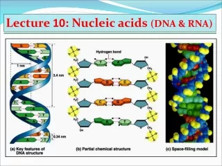
DNA Topology • Supercoiling or writhing of circular DNA is a result of the DNA being underwoundwith respect to the relaxed form of DNA • There are actually fewer turns in the DNA helix than would be expected given the natural pitch of DNA in solution (10.4 base pairs per turn) • When a linear DNA is free in solution it assumes a pitch which contains 10.4 base pairs per turn • This is less tightly wound than the 10.0 base pairs per turn in the Watson and Crick B-form DNA
DNA that is underwound is referred to as negatively supercoiled • The helices wind about each other in a right-handed path in space • DNA that is overwound will relax and become a positively supercoiled DNA helix • Positively coiled DNA has its DNA helices wound around each other in a left-handed path in space
Linking number - # times would have to pass cccDNA strand through the other to entirely separate the strands and not break any covalent bonds • Twist - # times one strand completely wraps (# helical turns) around the other strand • Writhe – when long axis of double helix crosses over itself (causes torsional stress)
Linking Defined • Linking number, Lk, is the total number of times one strand of the DNA helix is linked with the other in a covalently closed circular molecule
The linking number is only defined for covalently closed DNA and its value is fixed as long as the molecule remains covalently closed. • The linking number does not change whether the covalently closed circle is forced to lie in a plane in a stressed conformation or whether it is allowed to supercoil about itself freely in space. • The linking number of a circular DNA can only be changed by breaking a phosphodiester bond in one of the two strands, allowing the intact strand to pass through the broken strand and then rejoining the broken strand. • Lkis always an integer since two strands must always be wound about each other an integral number of times upon closure.
DNA tied up in knots • Metabolic events involving unwinding impose great stress on the DNA because of the constraints inherent in the double helix • There is an absolute requirement for the correct topological tension in the DNA (super-helical density) in order for genes to be regulated and expressed normally • For example, DNA must be unwound for replication and transcription Figure from Rasika Harshey’s lab at UT Austin showing an enhancer protein (red) bound to the DNA in a specific interwrapped topology that is called a transposition synapse.www.icmb.utexas.edu/.../47_Topology_summary.jpg
Knots, Twists, Writhe and Supercoiling • Circular DNA chromosomes, from viruses for instance, exist in a highly compact or folded conformation
Twist • The linking number of a covalently closed circular DNA can be resolved into two components called the twists, Tw and the writhes, Wr. • Lk = Tw + Wr • The twists are the number of times that the two strands are twisted about each other • The length and pitch of DNA in solution determine the twist. [Tw = Length (bp)/Pitch (bp/turn)]
Writhe • Writhe is the number of times that the DNA helix is coiled about itself in three-dimensional space • The twist and the linking number, determine the value of the writhe that forces the DNA to assume a contorted path is space. [Wr = Lk - Tw ]
Unlike the Twist and the Linking number, the writhe of DNA only depends on the path the helix axis takes in space, not on the fact that the DNA has two strands • If the path of the DNA is in a plane, the Wr is always zero • If the path of the DNA helix were on the surface of a sphere (like the seams of a tennis ball or base ball) then the total Writhe can also be shown to be zero
Molecules that differ by one unit in linking number can be separated by electrophoresis in agarose due to the difference in their writhe (that is due to difference in folding). • The variation in linking number is reflected in a difference in the writhe. • The variation in writhe is subsequently reflected in the state of compaction of the DNA molecule.
Interwound Toroidal Writhe of supercoiled DNA
Negative vs. Positive Supercoiling • Right handed supercoiling = negative supercoiling (underwinding) • Left handed supercoiling = positive supercoiling • Relaxed state is with no bends • DNA must be constrained: plasmid DNA or by proteins • Unraveling the DNA at one position changes the superhelicity
Relaxed Supertwisted
Ability of Uracil To Form Stable Base Pairs Enhances RNA’s Ability To Form Stem-loop Structures
Histone Variants • Alter nucleosome function • H2A.z often found in areas with transcribed regions of DNA • prevents nucleosome from forming repressive structures that would inhibit access of RNA polymerase • Mark areas of chromatin with alternate functions • CENP-A replaces H3 • Associated with nucleosomes that contain centromeric DNA • Has longer N-terminal tail that may function to increase binding sites available for kinetochore protein binding
more peripheral more central Unwrapping of DNA from nucleosome allows DNA-binding proteins access to their binding sites • Many DNA-binding proteins require histone-free DNA • DNA-histone interactions dynamic: unwrapping is spontaneous and intermittent • Accessibility to binding protein sites dependent on location in nucleosomal DNA • more central sites less accessible than those near the ends decreasing probability of protein binding and hence regulating transcriptional activity
Nucleosome remodeling complexes • Alter stability of DNA-histone interaction to increase accessibility of DNA • Change nucleosome location • Require ATP • 3 mechanisms: • Slide histone octamer along DNA • Transfer histone octamer to another DNA • Remodel to increase access to DNA
DNA-binding protein dependent nucleosome positioning • Nucleosomes are sometimes specifically positioned • Keeps DNA-binding protein site in linker region (hence accessible) • Can be directed by DNA-binding proteins or by specific sequences • Usually involves competition between nucleosomes and binding proteins • If proteins are positioned such that less than 147 bp exists between them, nucleosomes cannot associate
Positioning can be inhibitory • Some proteins can bind to DNA and a nucleosome • By putting a tightly bound binding protein next to a nucleosome, additional nucleosomes will assemble immediately adjacent to the protein preferentially
DNA sequences can direct positioning • DNA sequences that position nucleosomes are A-T or G-C rich because DNA is bent in nucleosomes • By alternating A-T or G-C rich sequences, can change the position in which the minor groove faces the histone octamer • These sequences are rare
Majority of nucleosomes are not positioned • Tightly positioned nucleosomes are usually associated with areas for transcription initiation • Positioned nucleosomes can prevent or enhance access to DNA sequences needed for binding protein attachment
Modification of N-terminal tails • Results in increased or decreased affinity of nucleosome for DNA • Modifications include acetylation, methylation and phosphorylation • Combination of modifications may encode information for gene expression (positively or negatively
Acetylated nucleosomes are associated with actively transcribed areas because reduces the affinity of the nucleosome for DNA • Deacetylation associated with inactive transcription units • Phosphorylation also increases transcription • Like acetylation, phosphorylation reduces positive charge on histone proteins • Methylation represses transcription • Also affects ability of nucleosome array to form higher order structures
HAT Acetylation creates binding sites for bromo- and chromodomain protein binding
Chromatin remodeling complexes and histone modifying enzymes work together to make DNA more accessible
Distributive inheritance of old histones • Old histones have to be inherited to maintain histone modifications and appropriate gene expression • H3▪H4 tetramers are randomly transferred to new daughter strand, never put into soluble pool • H2A▪H2B dimers are put into pool and compete for association with H3▪H4 tetramers
Histone assembly requires chaperones • Assembly of nucleosome is not spontaneous • Chaperone proteins are needed to bring in free dimers and tetramers after replication fork has been passed • Chaperones are associated with PCNA, the sliding clamp protein of eukaryotic replication, immediately after PCNA is released by DNA polymerase
Nucleotides and primer:template junction are essential substrates for DNA synthesis