Download
1 / 39
430 likes | 611 Views
Introduction to Laser Safety. Examples of laser accidents. Overview of the eye. macula or macula lutea (yellow spot on the retina): allows color vision

E N D
Overview of the eye macula or macula lutea (yellow spot on the retina): allows color vision fovea: central spot of macula allowing for sharp central vision (necessary for reading, TV, driving, and any activity where visual detail is of primary importance
413 Picosecond pulses cause bleeding/latent viewing distortion Description: New frequency doubler didn't have AR coatings as requested. As person left room, beam hit eye corner and transmitted schlera and caused interocular bleeding. Resided at 2 wks and eye normal at 2 months. Person still complains at 8 yrs of floaters and vision that looks "like looking through a dirty window". Type:FD Nd:YAG Divergence:- Wavelength:1064 nm Energy/Power:MW/cm2+ Class:IV Pulse Rate:1 KHz Exposure Time:60 ps
165 Reflected beam caused vision loss Description: Professor from China removed eyewear to "see better" while doing an experiment with a crystal. Exposure produced retinal burn and permanent vision loss. He described seeing a white flash, central purple spot surrounded by yellow halo. No pain reported.
223 Retinal burn from beam off rear laser mirror Description: Student WITH EYEWEAR ON (and witness to verify) received exposure from the rear mirror of a "Continuium" YAG laser. The student was wearing Glendale Broadband (OD 4.0) eyewear; ANSI standard requires OD=6.0. Retinal burn resulted with permanent damage. Type:Nd:YAG Divergence:- Wavelength:532 nm Energy/Power:0.18/0.40 Class:- Pulse Rate:5 KHz Exposure Time:7 ns
356 Blurred vision from reflected exposure. Description: Student received reflected beam from plastic tool box lid from Ti-Sapphire laser. No eye protection worn. Student reported blurred vision and seeing black spots. He was installing a laser transport tube (beam safety tube). The student had not received laser safety training. At 1 month student still had blurry vision. Type: Ti-Sapphire Divergence:- Wavelength:800 nm Energy/Power:15 mJ Class:IV Pulse Rate:10 KHz Exposure Time:120 fs
312 Off-axis beam causes macular burn in left eye Description: Scientist bumped mirror mount in a complex optical array - causing a stray beam to go off-axis. When leaning over the table, he was struck in left eye by beam off lower array mirror. Exam confirmed macular lesion which he states disrupts vision. No eyewear worn and safety knowledge was limited. Type:Ti-Sapphire Divergence:- Wavelength:800 nm Energy/Power:6 mJ Class:IV Pulse Rate:3.3K KHz Exposure Time:50 ns
307 Backscatter from mirror causes hemorrhage and oveal blindspot Description: A 26 year old male Student aligning optics in a university chemistry research lab using a "chirped pulse" Titanium-Sapphire laser operating at 815 nm with 1.2 mJ pulse energy at 1 KHz. Each pulse was about 200 picoseconds. The laser beam backscattered off REAR SIDE of mirror (about 1% of total) caused a foveal retinal lesion with hemorrhage and blind spot in central vision. A retinal eye exam was done and confirmed the laser damage. The available laser protective eyewear was not worn.
283 Photophobia in right eye after beam misalignment Description: Received "flash" into eye during alignment where he looked back along the beam path to view reflection off laser face plate. Result caused photophobia with burning sensation. No retinal burns detected. Patient used sunglasses for photophobia. Type:HeNe Divergence:- Wavelength:633 nm Energy/Power:6 mW Class:- Pulse Rate:- Exposure Time:~0.25 sec
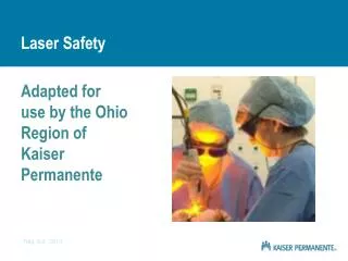
Airway Fire Description: During laser surgery on a patient’s vocal cords, the surgeon struck the endotracheal tube with a pulse from a CO2 laser. The tube, which carries oxygen to the patient and runs through his mouth to his lungs, was not made of laser-resistant material. Instead, it was made of polyvinyl chloride (combustible to both Nd:YAG and CO2 lasers). It caught fire and filled the man’s lungs with toxic smoke, causing burns. The patient did not survive the procedure. In general the anaesthesiologist has only six seconds to recognize that a tube has ignited and remove it before the fire peaks. Once ignited, the tubes are as hot as an oxygen lance used in welding. The flame can reach a length of 5 to 10 inches. Laser beam interaction with secondary materials is a known source of laser incidents. This sort of unplanned interaction is a danger one needs to think of beforehand.
Los Alamos Laser Accident Description: A postdoctoral employee received an eye exposure to spectral radiation from an 800 nm Class 4 laser beam. The extremely short pulse (100 fs) caused a 100-micron-diameter burn in the employee's retina. The accident occurred shortly after a mirror was removed from its mount and replaced with a corner cube during a realignment procedure. Although the beam had been blocked during several previous steps in the alignment, it was not blocked in this case. The employee was exposed to laser radiation from the corner cube mount when he leaned down to check the height of the mount. Neither of the two employees performing the alignment was wearing the appropriate laser eye protection. The system had two modes of operation: 10 Hz and 1,000 Hz. In addition, the researcher forgot that the part of the 800 nm beam he could see represented only 1-2% of the beam.
You have to ask yourselfcan this happen in our laboratories?
Dangers associated with the use of lasers • Beam hazards • eye damage • skin damage • Non-beam hazards • electrical hazards • toxic/carcinogenic laser dyes • hazardous gases (e.g. excimer lasers) • fire
Majority of injuries involve the eye and to lesser extend the skin Summary of reported laser accidents in the United States and their causes from 1964 to 1992
Majority of injuries during alignment, or no use or improper use of eyewear Summary of reported laser accidents in the United States and their causes from 1964 to 1992
Potential eye damage In general terms, in supra-threshold exposures the predominating mechanism is broadly related to the pulse duration of the exposure. Thus, in order of increasing pulse duration, the predominant effects in the following time domains are: • nanosecond and sub-nanosecond exposures, acoustic transients and non-linear effects • from 1 ms to several seconds, thermal effects • in excess of 10 s, photochemical effects.
Potential eye damage The biological damage caused by lasers is produced through thermal, acoustical and photochemical processes. Thermal effects are caused by a rise in temperature following absorption of laser energy. The severity of the damage is dependent upon several factors, including exposure duration, wavelength of the beam, energy of the beam, and the area and type of tissue exposed to the beam. Normal focusing by the eye results in an irradiance ampli-fication of roughly 100,000; therefore, a 1 mW/cm2 beam en-tering the eye will result in a 100 W/cm2 exposure at the retina. The most likely effect of intercepting a laser beam with the eye is a thermal burn which destroys the retinal tissue. Since retinal tissue does not regenerate, the damage is permanent.
Potential eye damage Acoustical effects result when laser pulses with a duration less than 10 microseconds induce a shock wave in the retinal tissue which causes a rupture of the tissue. This damage is perma-nent, as with a retinal burn. Acoustic damage is more destructive than a thermal burn. Acoustic damage usually affects a greater area of the retina, and the threshold energy for this effect is substantially lower. Beam exposure may also cause Photochemical effects when photons interact with tissue cells. A change in cell chemistry may result in damage or change to tissue. Photochemical effects depend strongly on wavelength. N.B. the severity of the damage depends strongly on whether it occurs by intrabeam exposure or scattered laser light
Skin hazards • In general terms, the skin can tolerate a great deal more exposure to laser beam energy than can the eye. • The biological effect of irradiation of skin by lasers operating in the visible and infra-red spectral regions may vary from a mild erythema to severe blisters. • An ashen charring is prevalent in tissues of high surface absorption following exposure to very short-pulsed, high-peak power lasers. • The pigmentation, ulceration, and scarring of the skin and damage of underlying organs may occur from extremely high irradiance. • In the wavelength range 1500 nm to 2600 nm, biological threshold studies indicate that the risk of skin injury follows a similar pattern to that of the eye.
Example of eye injury Experience has demonstrated that most laser injuries go unreported for 24–48 hours by the injured person. This is a critical time for treatment of the injury.
Retinal Burn A range of injuries induced with a Nd:YAG laser on a monkey retina. The white spots in the centre are thermal burns, i.e. coagulation of retinal layers. With larger energies, holes in the retina are produced which result either in bleeding into the vitreous (the gel-like substance which fills the centre of the eye ball), or the bleeding is contained in the layers of the retina, which results in functional loss in the affected area. Photograph courtesy of J. Zuclich, TASC Litton, TX, USA.
Eye Damage: FocusingRemember: Your Eyes Are Designed to Focus With safety rule Cornea Damage BAD Retina Damage WORSE
Laser classification Class 1 Lasers Lasers that are safe under reasonably foreseeable conditions of operation, including the use of optical instruments for intrabeam viewing. Class 1M Lasers Lasers emitting in the wavelength range from 302,5 nm to 4000 nm which are safe under reasonably foreseeable conditions of operation, but may be hazardous if the user employs optics within the beam.
Laser classification Class 2 Lasers Lasers that emit visible radiation in the wavelength range from 400 nm to 700 nm where eye protection is normally afforded by aversion responses, including the blink reflex. This reaction may be expected to provide adequate protection under reasonably foreseeable conditions of operation including the use of optical instruments for intrabeam viewing. Outside this wavelength range AEL ≤ AEL of a class 1 laser. Class 2M Lasers Like class 2 lasers, however, viewing of the output may be more hazardous if the user employs optics within the beam. Outside visible range AEL ≤ AEL of a class 1M laser.
Laser classification Class 3R Lasers Lasers that emit in the wavelength range from 302,5 nm to 106 nm where direct intrabeam viewing is potentially hazardous but the risk is lower than for Class 3B lasers. The accessible emission limit is within five times the AEL of Class 2 in the wavelength range from 400 nm to 700 nm and within five times the AEL of Class 1 for other wavelengths. Class 3B Lasers Lasers that are normally hazardous when direct intrabeam exposure occurs. Viewing diffuse reflections is normally safe.
Laser classification Class 4 Lasers Lasers that are also capable of producing hazardous diffuse reflections. They may cause skin injuries and could also constitute a fire hazard. Their use requires extreme caution
Retinal injury thresholds Health Physics October 2000, Volume 79, Number 4 At 10-12 seconds the threshold for a retinal injury is appr. 10-7 J/cm2 (i.e. 105 W/cm2). Because of the x 105 enhancement in the eye this value is elevated to 10-2 J/cm2 (i.e. 1010 W/cm2) on the retina. These exposure levels are further enhanced by self-focussing.
Exposure limits, Retinal injuryexample • A 4 % reflection from a 2.5 mJ laser pulse in a 2 mm beam, gives an exposure of (10-4 J)/(p x 0.12 cm2) = 3.2 10-3 J/cm2. • This exceeds the threshold value of the cornea of about 10-7 J/cm2 by a factor of 3.2 104. • To be adequately protected against this exposure, protective eyewear must have an optical density (OD) of at least log10(3.2 104) = 4.5
Some common unsafe practices: preventable laser accidents • Not wearing protective eyewear during alignment procedures • Not wearing protective eyewear in the laser control area • Misaligned optics and upwardly directed beams • Equipment malfunction • Improper methods of handling high voltage • Available eye protection not used • Intentional exposure of unprotected personnel • Lack of protection from non-beam hazards
Some common unsafe practices or preventable laser accidents • Failure to follow (Laser) Safety Instructions • Bypassing of interlocks, door and laser housing • Insertion of reflective materials into beam paths • Lack of pre-planning • Turning on power supply accidentally • Operating unfamiliar equipment • Wearing the wrong eyewear
Guidelines to help preventaccidents during alignment • No unauthorized personnel will be in the room or area. • Laser protective eyewear will be worn. • The individual who moves or places an optical component on an optical table is responsible for identifying and terminating each and every stray beam coming from that component. • To reduce accidental reflections, watches and reflective jewellery should be taken off before any alignment activities begin. • Beam blocks must be used and must be secured. • When the beam is directed out of the horizontal plane, it must be clearly marked.
Guidelines to help preventaccidents during alignment • The lowest possible/practical power must be used during alignments. • Have beam paths that differ from the eye level when standing or sitting. Do not use paths that tempts one to bend down and look into the beam. • All laser users must receive an introduction to the laser area by an authorised laser user of that area
Responsibilities The department is responsible for the safety of its employees and exercises its responsibility by providing guidelines and periodic control by designated safety personnel. We all have a personal responsibility to make sure that our working conditions and working habits are safe and in accordance with the guidelines.
Acknowledgement / References • This presentation is inspired by a similar presentation on Laser Safety by the Molecular & Laser Physics Group of the Radboud University, Nijmegen NL. • Laser incidents taken from laser accident database of Rockwell Laser Industries, Inc. (http://www.rli.com/resources/accident.asp) • Certain descriptions and numbers are taken from the international standard: IEC 60825-1 Edition 1.2. • The model of the eye is provided by the National Eye Institute, USA.
Aqueous flare = the presence of floating particles in the fluid of the anterior chamber of the eye Cataract = opacity or reduction in clarity of the lens of the eye Erythema = redness of the skin caused by dilatation and congestion of the capillaries, often a sign of inflammation or infection Fovea centralis = a small depression near the center of the retina, constituting the area of most acute vision Inflammation = A localized protective reaction of tissue to irritation, injury, or infection, characterized by pain, redness, swelling, and sometimes loss of function Lesion = a localized pathological change in a bodily organ or tissue Macula letua (yellow spot) = A minute yellowish area containing the forea centralis located near the center of the retina of the eye at which visual perception is most acute. Macular lesion = Loss of central vision Photokeratitis = Inflammation of the cornea produced by ultraviolet radiation Photophpobia = an abnormal sensitivity to or intolerance of light, especially by the eyes, as may be caused by eye inflammation, lack of pigmentation in the iris, or various diseases Schlera = outer hard white coat (cover) of the eyeball