Download
1 / 12
120 likes | 486 Views
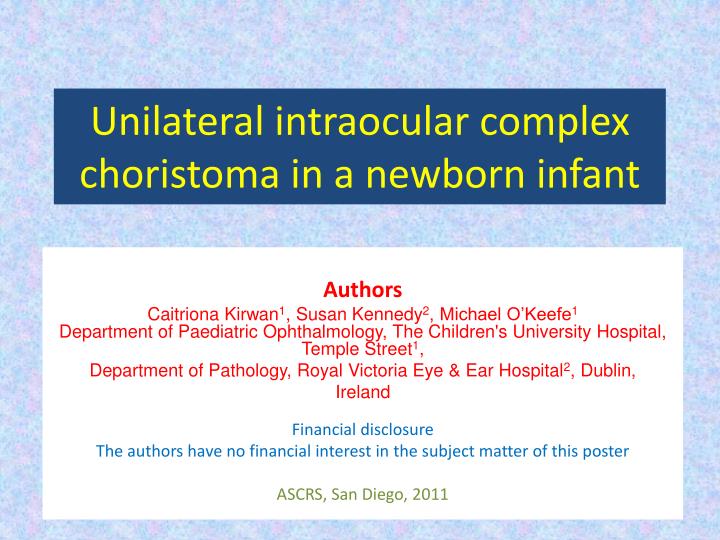
Unilateral intraocular complex choristoma in a newborn infant. Authors Caitriona Kirwan 1 , Susan Kennedy 2 , Michael O’Keefe 1 Department of Paediatric Ophthalmology, The Children's University Hospital, Temple Street 1 , Department of Pathology, Royal Victoria Eye & Ear Hospital 2 , Dublin,

E N D
Unilateral intraocular complex choristoma in a newborn infant Authors Caitriona Kirwan1, Susan Kennedy2, Michael O’Keefe1Department of Paediatric Ophthalmology, The Children's University Hospital, Temple Street1, Department of Pathology, Royal Victoria Eye & Ear Hospital2, Dublin, Ireland Financial disclosure The authors have no financial interest in the subject matter of this poster ASCRS, San Diego, 2011
Background • A female infant was born to healthy, unrelated parents following an uneventful pregnancy and delivery • At birth she was found to have a large mass protruding from the left orbit, completely replacing the normal globe • The mass was well circumscribed and moved partially on movement of the other eye indicating some attachment to extra-ocular muscles. However, normal eye structures were not apparent. The upper and lower eyelids were stretched significantly to accommodate the large mass • Systems review revealed no other abnormalities
Normal eye structures not evident Transillumination
The child was brought to the operating room where examination was performed Exploration of the mass posterior to the equator revealed the presence of extraocular muscles Rectus muscle
Following removal of the mass – the contour of the eyelids was more evident. Both the upper and lower lids had in effect ‘cicatricial’ ectropions with extremely tight skin and absent fornices The width of the palpebral aperture from nasal to temporal extremities was increased compared to the fellow eye A conformer was placed in the socket and a lateral tarsorrhaphy was performed
Cornea replaced by opaque yellow plaque • The mass measured 30mm in length and 24×25mm in width • Differential diagnosis: • Congenital cystic eye • Cystic teratoma ←30mm→ ←25mm → ←24mm→ • Histological analysis was performed Optic nerve
Histology • Cystic, partially calcified lesion • Skin & subcutaneous tissue over sclera • Anteriorly complex solid/cystic mass: epithelial inclusion cyst surrounded by mature fat tissue, mesenchymal tissue including striated muscle and neural tissue with ependymal-like cystic structures • Posteriorly: small amount of compressed intraocular tissue – choroid, retina & poorly formed anterior segment structures, RPE & ciliary body
Histology • Retina: small areas of intra-retinal calcification, retinal dysplasia & well formed rosettes & nodule of vascularised fibrous tissue & adipose tissue • No evidence of cornea, anterior chamber or angle structures or lens • Optic nerve histologically normal • Diagnosis: Extensive anterior intraocular complex choristoma with hypoplastic intraocular tissues
Congenital Cystic Eye • Rare ocular and orbital malformation describing an intraorbital cavity lined by neuroglial tissue • Primary developmental abnormality of the globe caused by an invaginational arrest of the primary optic vesicle between the 2 & 7mm stages of fetal development • ~30 reported cases in literature • Chaudhry et al, IntOphthal 2007 • Gupta et al, BMC Ophthal 2003 • Frequently associated systemic abnormalites • Anophthalmia
Teratoma • Rare congenital choristoma arising from all 3 germ layers; ectoderm, mesoderm & endoderm • Orbital teratoma – rare congenital tumour • 2:1 female preponderance • Normal fellow eye • Rare cases of malignant transformation • 2 previous reports of teratoma arsing within the globe • Leventer et al. AJO 2001 • Kivela et al. Ophthalmology 1993 • Pathogenesis: • Circulating pluripotential stem cells carried haematogenously to the orbit are incorporated within the eye during closure of the embryonic fissure
Choristoma • Congenital proliferation of histologically mature tissue elements not normally present at the site of occurrence eg limbaldermoid, dermolipoma • Composed of tissue arising from1 or 2 germ layers • Example: Osseous choristoma Lacrimal gland choristoma Phakomatous choristoma of eyelid • First reported case of a true complex intraocular choristoma
Conclusion • Extremely rare case • Cosmesis an important issue • Multiple conformers inserted • Skin graft to lower lid • Latest follow up: • 4 years old • Good cosmesis • Prosthesis fits securely in socket