Download
1 / 42
420 likes | 702 Views
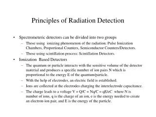
Introduction. Increasing use for head and neck cancerCombined or as single modalityOutline basic principles, radiobiologyGeneral treatment approachCommon complications. Radiation Physics. Basis ionizing particles interact with cellular moleculesRelies on transfer of energy created by secondary charged particles (usually electrons)Break chemical bondsExternal beam vs. BrachytherapyRadiant energy is discrete yet random.

E N D
1. Principles of Radiation Oncology Michael Underbrink, MD
Anna Pou, MD
2. Introduction Increasing use for head and neck cancer
Combined or as single modality
Outline basic principles, radiobiology
General treatment approach
Common complications
3. Radiation Physics Basis � ionizing particles interact with cellular molecules
Relies on transfer of energy created by secondary charged particles (usually electrons)
Break chemical bonds
External beam vs. Brachytherapy
Radiant energy is discrete yet random
4. External Beam Irradiation Dual-energy linear accelerators generate:
Low energy megavoltage x-rays (4-6 MeV)
High energy x-rays (15-20 MeV)
Photon energy
Particle Radiation (electrons, protons, neutrons)
Photon therapy advantages
Skin sparing, penetration, beam uniformity
Head and Neck sites � 4-6 MeV x-ray or Co60 gamma ray radiation
5. External Beam Irradiation
6. Brachytherapy Radioactive source in direct contact with tumor
Interstitial implants, intracavitary implants or surface molds
Greater deliverable dose
Continuous low dose rate
Advantage for hypoxic or slow proliferators
Shorter treatment times
7. Brachytherapy Limitations
Tumor must be accessible
Well-demarcated
Cannot be only modality for tumors with high risk of regional lymph node metastasis
8. Brachytherapy
9. Radiobiology Ionizing radiation ejects an electron from a target molecule
Distributed randomly within cell
Double-strand DNA breaks � lethal
Cell death: no longer able to undergo unlimited cell division
Direct vs. Indirect injury (free radicals � O2)
Inadequate cellular repair mechanisms implied
10. Radiobiology
11. Radiobiology Random cell death
Deposition of energy & injury is random event
Same proportion of cells is damaged per dose
100 to 10 cell reduction = 106 to 105 cell reduction
Larger tumors require more radiation
105 cells = nonpalpable
Applies to normal tissue also
Therapeutic advantage � 4 R�s of radiobiology
12. 4 R�s of radiation biology Repair of cellular damage
Reoxygenation of the tumor
Redistribution within the cell cycle
Repopulation of cells
13. Repair of sublethal injury Sublethal injury � cells exposed to sparse ionization fields, can be repaired
Killing requires greater total dose when given in several fractions
Most tissue repair in 3 hours, up to 24 hours
Allows repair of injured normal tissue, potential therapeutic advantage over tumor cells
Radioresistance � melanoma?
14. Reoxygenation Oxygen stabilizes free radicals
Hypoxic cells require more radiation to kill
Hypoxic tumor areas
Temporary vessel constriction from mass
Outgrow blood supply, capillary collapse
Tumor shrinkage decreases hypoxic areas
Reinforces fractionated dosing
Hypoxic cell radiosensitizers, selective chemo
15. Reoxygenation
16. Redistribution Cell cycle position sensitive cells
S phase � radioresistant
G2 phase delay = increased radioresistance
RAD9 gene mutation � radiosensitive yeast
H-ras and c-myc oncogenes - G2 delay
Fractionated XRT redistributes cells
Rapid cycling cells more sensitive (mucosa, skin)
Slow cyclers (connective tissue, brain) spared
17. Redistribution
18. Repopulation Increased regeneration of surviving fraction
Rapidly proliferating tumors regenerate faster
Determines length and timing of therapy course
Regeneration (tumor) vs. Recuperation (normal)
Reason for accelerated treatment schedules
Reason against:
Treatment delay
Protracted XRT, split course XRT (designed delay)
19. Repopulation
20. Dose-Response Relations Control probability variables
Tumor size
XRT dose
Favorable response curves
Small, well-vascularized tumors
Homogeneous tumors
Unfavorable response curves
Large, bulky tumors (hypoxia)
Heterogeneous, variable cell numbers
Normal tissue injury risk increases with XRT dose (size of tumor)
21. Dose-Response Relations
22. Fractionation
23. Fractionation Schedules Conventional
1.8 to 2.0 Gy given 5 times/week
Total of 6 to 8 weeks
Effort to minimize late complications
Accelerated fractionation
1.8 to 2.0 Gy given bid/tid
Similar total dose (less treatment time)
Minimize tumor repopulation (increase local control)
Tolerable acute complications (increased)
24. Fractionation Schedules Hyperfractionation
1.0 to 1.2 Gy bid/tid, 5 times/week
Similar total treatment time (increased total dose)
Increases total dose
Potentially increases local control
Same rates of late complications
Increased acute reactions
25. Treatment Principles Size and location of primary
Presence/absence and extent/incidence of regional or distant metastasis
General condition of patient
Early stage cancers
Surgery alone = XRT alone
Treatment choice depends on functional deficits
Late stage � usually combination of treatments
26. Treatment Principles Surgical salvage of primary radiation failures is better than radiation salvage of surgical failure
Explains rationale behind organ preservation strategies
XRT tumor cell killing is exponential function
Dose required for tumor control is proportional to the logarithm of the number of viable cells in the tumor
27. Shrinking field technique Initial dose = 45 to 50 Gy (4.5 to 5.0 weeks)
Given through large portals
Covers areas of possible regional metastasis and primary
Second dose = 15 to 25 Gy (1.5 to 2.5 weeks)
Boost field (gross tumor and small margin)
Total dose of 60 to 75 Gy in 6 to 7.5 weeks
Boost dose = 10 to 15 Gy
Massive tumors
Second field reduction at 60 to 65 Gy
Total of 7 to 8 weeks
28. Shrinking field technique
29. Combined Modalities Surgery and XRT complement each other
Surgery � removes gross tumor (bulky tumors are more difficult to control with XRT)
XRT � effective for microscopic disease, better with exophytic tumors than ulcerative ones (Surgical failures may leave subclinical disease)
Combining treatments counteracts limitations
Pre or Post-operative XRT
30. Preoperative XRT Advantages
Unresectable lesions may become resectable
Extent of surgical resection diminished
Smaller treatment portals
Microscopic disease more radiosensitive (blood supply)
Decreased risk of distant metastasis from surgical manipulation?
Disadvantages
Decreased wound healing
Decreased safe dose (45 Gy in 4.5 weeks eradicates subclinical disease in 85% to 90% of patients)
31. Postoperative XRT Advantages
Better surgical staging
Greater dose can be given safely (60 to 65 Gy in 6 to 7 weeks)
Total dose can be based on residual tumor burden
Surgical resection is easier
Tissue heals better
Disadvantages
Distant metastasis by manipulation?
Delay in postoperative treatment if healing problems (poorer results if delayed more than 6 weeks)
32. Complications Acute Tissue Reactions
Late Tissue Reactions
33. Acute Toxicity Time onset depends on cell cycling time
Mucosal reactions � 2nd week of XRT
Skin reactions � 5th week
Generally subside several weeks after completion of treatment
RTOG � acute toxicity <90 days from start of treatment (epithelial surfaces generally heal within 20 to 40 days from stoppage of treatment)
34. Acute Toxicity Mucositis � intensity-limiting side effect for aggressive schedules
Accelerated fractionation � increase acute toxicities
Conventional fractionation conservatively emphasized maximum tolerated dose is limited by late not acute tissue injury
35. Acute Toxicity
36. Late Toxicity Injury tends to be permanent
Cells with low turnover (fibroblasts, neurons)
Develop within months to years
Xerostomia, dental caries, fibrosis, soft-tissue necrosis, nerve tissue damage
Most common - xerostomia
37. Late Toxicity
38. Late Toxicity Xerostomia
Injury to serous acinar cells
May have partial recovery
Results in dental caries (in or outside of fields)
Soft tissue necrosis
Mucosal ulceration, damage to vascular connective tissue
Can result in osteo-/chondroradionecrosis
39. Late Toxicity
40. Late Toxicity Fibrosis
Serious problem, total dose limiting factor
Woody skin texture � most severe
Large daily fractions increase risk
Ocular � cataracts, optic neuropathy, retinopathy
Otologic � serous otitis media (nasopharynx, SNHL (ear treatments)
41. Late Toxicity
42. Late Toxicity Central Nervous System
Devastating to patients
Myelopathy (30 Gy in 25 fractions)
Electric shock from cervical spine flexion (Lhermitte sign)
Transverse myelitis (50 to 60 Gy)
Somnolence syndrome (months after therapy)
Lethargy, nausea, headache, CN palsies, ataxia
Self-limiting, transient
Brain necrosis (65 to 70 Gy) � permanent
43. Conclusions XRT key role in treatment of H&N cancer
Fundamentals of radiation physics and radiobiology explain rationale behind treatment schedules and complications
Basic knowledge important with regard to patient counseling