Download

1 / 62
780 likes | 1.36k Views
Nephrology 1 Pathophysiology of upper tract obstruction. Euan Green Mr Brough. 40 yr old man Referred by GP Vague abdominal pain USS shows solitary left kidney with hydronephrosis Seen in clinic and asymptomatic Wants to know “What’s up doc?”. Does this mean my kidney is blocked?. No!

E N D
Nephrology 1Pathophysiology of upper tract obstruction Euan Green Mr Brough
40 yr old man • Referred by GP • Vague abdominal pain • USS shows solitary left kidney with hydronephrosis • Seen in clinic and asymptomatic • Wants to know “What’s up doc?”
Does this mean my kidney is blocked? • No! • Hydronephrosis = dilatation of renal pelvis or calyces with or without obstruction • Hydroureteronephrosis = above + dilated ureter • Obstruction = obstruction to the flow of urine
What’s the cause? • Congenital or acquired • Intra-luminal, in the lumen wall or extraluminal • Complete or incomplete
What do you do next? • U&E? • CT? • Renogram? Na+ 140 K+ 4.2 Urea 5.0 Creat 70
What do you do next? • U&E? • CT? • Renogram?
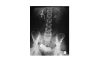
What is the diagnosis? • Extrarenal pelvis
25 yr old woman • Sudden onset severe right loin pain • Colicky • Vomiting • Dipstick haematuria • Comes to A&E • Seen by SHO who arranges a CT Urogram
Your SHO asks you “what will happen? Will the kidney just fill up and then burst?” • Triphasic response to upper tract obstruction • Different for unilateral and bilateral obstruction
The contralateral kidney • Compensatory Growth • Response proportional to degree of injury • Initial vasoconstriction, subsequent vasodilation • Hypertrophy • Increased blood flow and GFR • Compensatory growth is age dependent • The number of nephrons remains constant • Increase in proximal tubular length due to increase in cell size
75 yr old man • Previously completely well • Nocturnal enuresis for the last 6 weeks • Tired • Pitting ankle oedema • BP 180/100 • Large abdominal mass
What is the likely diagnosis? • High pressure Chronic retention of urine • Initial management? • U&E • Creatinine 1500, Urea 43, K+ 6.8 • Admit as an emergency • Catheterise • 3.5 Litre residual • ECG & Treat K+ • CaGluconate, insulin dextrose, fluids, salbutamol • If K+ still high with ECG changes? • Dialysis • USS urinary tract
The patient wants to know: • What has happened to his kidneys • What to expect over the next few days • Whether his kidney function will recover
Bilateral obstruction • Similar to unilateral upper tract obstruction • Less pronounced rise in blood flow initially • Less afferent vasodilation • Lasts 90 mins • More substantial decline in blood flow after • Greater vasoconstriction (thought to be due to no renal clearance of vasoconstricters from other kidney) • Renal pelvic and ureteric pressures remain raised for longer, approaching pre-obstruction pressure at 24 hrs • No other side to compensate
Macroscopic effects on kidney • Dilatation of pelvis/calyxes – hydronephrosis • Dilation of ureter • Flattened papillae (42hrs) • Parenchymal oedema (7 days) • Cortical parenchymal thinning (21 days)
Microscopic effects • 42 hrs – lymphatic dilatation, interstitial oedema • 7 days – collecting duct, tubular dilation, widening of Bowman’s space, tubular basement membrane thickening • 9 days – papillary tip necrosis and inflammatory cell infiltrate • 16 days – interstitial fibrosis • 3 weeks – tubular loss, fibroblast growth, collagen deposition • 6 weeks – Widespread tubular atrophy and fibrosis • Apoptosis is the principle mechanism of cell loss
Effect on tubular function • Down-regulation of aquaporin channels impairs concentrating ability • Some down-regulation of active sodium transporters. In addition fluid overload stimulates ANP secretion encouraging natriuresis • Reduction in GFR and down-regulation of Na+/K+ ATPase transporters reduces K+ excretion • Down-regulation of active H+ transporters results in a relative failure of H+ ion excretion • In unilateral obstruction the other kidney can compensate
Post-obstructive diuresis • Rare after relief of unilateral obstruction • Typically a physiological response to retained solutes (urea, sodium) and water • Pathological component due to tubular defects as mentioned, in particular the downregulation of aquaporin channels resulting in reduced sensitivity to ADH and the obliteration of the concentration gradient around the loop of Henle • Can be due to excess fluid replacement • Some patients develop hyponatraemia and hyperkalaemia due to tubular resistance to aldosterone
Post-obstructive diuresis • Those at increased risk: • Hypertension • Oedema • CCF • Long standing obstruction • Clinical uraemia • 20% have a urine output >4L in 24hrs • 5-10% require IV fluids • ~1% develop long term salt loss/diuresis
Return of renal function • Degree function return difficult to predict, relates to degree of obstruction, duration and prior function • Dog experiments have been carried out: • 7 days: full functional recovery • 14 days: 70% recovery • 28days: 30% recovery • 6 weeks: no functional recovery • In humans return of function has been noted after 150 days • Difficult to predict • 2 phases of recovery: • First 2 weeks – recovery in tubular function • Next 10 weeks – recovery in GFR
21 year old woman comes to the haematuria clinic • Left sided loin pain after nights out in the pub • Occasional haematuria • Otherwise fit and well • Flexible cystoscopy normal • Young so she gets an MRI rather than a CT
She wants to know what the diagnosis is? • Left PUJ obstruction • 1:500-1000 • 50% present in adult life, 50% as a child • Male 2:1 Female, Left > Right • Associated with other abnormalities in 50% • 10% bilateral • VUR • Horseshoe/renal duplication/ectopia • Due to crossing vessel/poor canalisation during formation/abnormal insertion into renal pelvis/ smooth muscle mal-development causing atonic segment
Right – 18% Left – 82% • What are you going to do next? • Confirm obstruction and assess function in that kidney. i.e. do a renogram • She wants to know: “how can this be treated?” • Conservative • Stent • Endopyelotomy • Pyeloplasty
Treatment options in PUJO • Conservative Mx • Suitable if asymptomatic and >40% relative function and unilateral • Need monitoring • Asymptomatic, non-functioning units • Stent • Palliation for the unfit • Management of the acute presentation • ?Diagnostic role
Treatment options in PUJO • Endopyelotomy • Ureteroscopy + laser/knife (60-85% success) • Acucise Device (Retrograde balloon catheter + cutting wire) (65-80% success) • Percutaneous endopyelotomy (80% success) • See NICE guidance Dec 2009 • Various definitions of success – all relatively short term success
Treatment options in PUJO • Pyeloplasty (>90% success) • Open • Laparoscopic • Robotic • Aim of surgery is to restore normal urine flow • Anastomosis has to be watertight and funnelled into the ureter
Anderson-Hynes Pyeloplasty • Excise PUJ • Spatulate the ureter • Narrow pelvis defect if large • Anastomose spatulated ureter to pelvis • Usually over a stent
Culp-DeWeerd Spiral flap • Suited to those with a long stricture • Open a spiral flap of renal pelvis • Rotate down and use flap to augment the width of the stenosed ureter
Foley V-Y Pyeloplasty • Useful for high ureteric insertion into pelvis • V-shaped flap from renal pelvis • Inserted into Y-shaped defect opened over stricture
Ureterocalicostomy • Suitable for revision surgery or renal abnormalities that prevent other options • Anastomose ureter to a lower pole calyx • Requires lower pole partial nephrectomy to reduce risk of stenosis
Treatment options in PUJO • Most now managed with laparoscopic pyeloplasty • Poorly functioning kidneys • <15-20% • Can consider nephrectomy
50 yr old man • Presents with loin pain • Intermittent • Can be severe • GP arranges USS which shows hydronephrosis • A medical student in clinic asks “What types of imaging can be used to demonstrate obstruction?”
Imaging for obstruction • USS • Can show dilatation (ie hydronephrosis) • False +ve • Excess flow eg Diuresis • Anatomy eg Extrarenal pelvis, Cysts • False –ve • Too little flow eg dehydration • Operator dependant • Can use doppler renal resistive index • >0.7 suggests obstruction, ~0.6 normal • (Peak systolic velocity-peak diastolic velocity) / Peak systolic velocity
Imaging for obstruction • IVU • Dynamic test • Functional information • Complete vs partial • Level of obstruction • Time consuming in obstructed patients
Imaging for obstruction • CT with or without contrast • Cheap • Quick • Good at identifying causes both intrinsic and extrinsic • Comparatively high radiation dose
Imaging for obstruction • MRI • Can identify hydronephrosis • Can’t detect stones • No radiation • Useful in the pregnant patient
Imaging for obstruction • Renogram • A study of the uptake, transit and elimination by the kidney of an intravenous dose of a radionucleotide • Gives drainage and relative function • Limited anatomical information • Use of diuretic improves discrimination between obstructed and non-obstructed
DOSE % 0 1 2 3 12 Renal 8 Bladder 4 TIME (minutes) 10 20 30 Renography • 3 phases • Vascular phase, represents uptake • Transit phase, represents transit through kidney • Elimination phase, excretion from the kidney and expulsion down the ureter
O’Reilly Curves • F+20 renogram
Back to the patient • What does this show? • What next? • F-15 Renogram • What if it’s still equivocal? • Whittaker’s test
Whittaker’s test • A test to help differentiate in those with equivocal obstruction or poor function where renogram unhelpful • Quite invasive • Nephrostomy in affected kidney • Catheterise • Patient prone in fluoroscopy • Infuse contrast/saline via nephrostomy at 10mls/min • Measure pressure in kidney and bladder and subtract to get the difference • <15 cm H20 – unobstructed • 15-22 cm H20 – equivocal • >22 cm H20 - obstructed