Download
1 / 42
420 likes | 514 Views
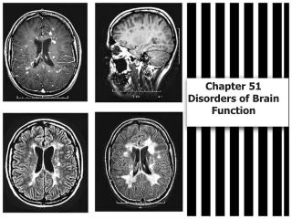
Bi 1 Session 4 Monday, April 3, 2006 What is a Brain? Introduction to neuronal circuits, neurons, synapses, non-invasive imaging, and multi-electrode recording. Hemispheres. MRI imaging. Regions. PET imaging. Connections. 2-deoxyglucose labelling. Neurons. Multi-cell recordings.

E N D
Bi 1 Session 4 Monday, April 3, 2006 What is a Brain? Introduction to neuronal circuits, neurons, synapses, non-invasive imaging, and multi-electrode recording
Hemispheres MRI imaging Regions PET imaging Connections 2-deoxyglucose labelling Neurons Multi-cell recordings Synapses Single-cell recordings Organization of the Brain Electrical and chemical events
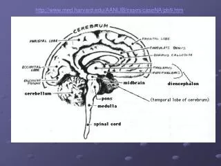
Brain Spinal cord Front Back The Central Nervous System:
Function is localized to each hemisphere Roger Sperry of Caltech (Nobel Prize,1981) Sperry investigated patients whose corpus callosum had been cut to stop intractable epilepsy. The surgeries were performed by Dr. Joseph Bogen (1926-2005), a neurosurgeon.
Spatial abilities,Face recognition,Visual imagery, Music A split-brain patient fixates on the dot in the middle of a screen. Then a picture of a spoon is flashed to the right of the dot. Language, Math,Logic The visual information about the spoon crosses via the optic nerve and travels to the LEFT HEMISPHERE. The person correctly identifies the spoon verbally.
Language, Math,Logic Spatial abilities,Face recognition,Visual imagery, Music Now the picture of a spoon is flashed to the left of the dot. Now the visual information travels to the RIGHT HEMISPHERE. Now if the subject is asked to identify the picture, he reports seeing nothing. But, when this same subject is asked to pick out an object using only the LEFT hand, he correctly picks out the spoon. This is because touch information from the left hand crosses over to the right hemisphere - the side that "saw" the spoon. However, if he is again asked to identify the object verbally, even when it is in his hand, he cannot do so because the right hemisphere cannot "talk." So, the right hemisphere is not stupid, it just has little ability for language - it is "non-verbal."
Function is often localized to specific brain regions acetylcholine (nicotine) and dopamine Function is often localized to specific brain regions front back Front Back memory (hippocampus)
A typical pathway: sensation of pain and the reaction to pain
Spinal reflexes, such as the knee-jerk, involve just two neurons. sensory neuron motor neuron the sensory neuron acts like a strain gauge wrapped around a special muscle fiber.
direction of information flow Postsynaptic Presynaptic neuron neuron Greek, “axis” Greek, “tree” Parts of two neurons Inhibitory terminal Excitatory terminal presynaptic terminal axon cell body dendrites presynaptic terminal nucleus postsynaptic dendrite synaptic cleft Nestler Figure 2-2 (rotated)
Greek, “connection, junction” The synapse is a point of information processing presynaptic neuron postsynaptic neuron Nestler Box 2 - 3 Figure A An adult human brain contains ~ 1011 neurons, and each of thesemight receive 103 synapses apiece, for a total of 1014 synapses. Most of these synapses form during the first 2 yr of life. Thus 1014synapses/108 s = 106 synapses/s form in a fetus and infant!
direction of information flow Chemistry is a language of the nervous system, for instance at synapses cytosol synaptic cleft cytosol receptor presynaptic terminal postsynaptic dendrite receptor transmitter molecules receptor
Electricity is a language of the nervous system Nestler Figure 3-1B Modified from Nestler Figure 3-1B
V Intracellular recording with sharp glass electrodes 1. Responses to artificially applied current pulses http://info.med.yale.edu/neurobio/mccormick/movies/fs_ctx1.mpg http://info.med.yale.edu/neurobio/mccormick/movies/fs_ctx1.avi Same data; choice of 3 formats. Media player required http://info.med.yale.edu/neurobio/mccormick/movies/fs_ctx1.mov (The spikes in these examples are about 100 mV in amplitude)
V Intracellular recording with sharp glass electrodes 2. Artificially applied acetylcholine acts on nicotinic acetylcholine receptors to produce currents http://info.med.yale.edu/neurobio/mccormick/movies/ach_fin.mpg http://info.med.yale.edu/neurobio/mccormick/movies/ach_fin.avi Same data; choice of 3 formats. Media player required http://info.med.yale.edu/neurobio/mccormick/movies/ach_fin.mov (The spikes in these examples are about 100 mV in amplitude)
V Intracellular recording with sharp glass electrodes 3. A cell is receiving stimuli from other cells, not from the experimenter http://info.med.yale.edu/neurobio/mccormick/movies/rly_exp.mpg Same data; choice of 3 formats. Media player required http://info.med.yale.edu/neurobio/mccormick/movies/rly_exp.avi http://info.med.yale.edu/neurobio/mccormick/movies/rly_exp.mov (The spikes in these examples are about 100 mV in amplitude)
2-deoxyglucose can label thousands or millions of active cells at once cell activity glucose glucose phosphates metabolic products, ATP cell activity 2-deoxyglucose (radiolabelled) 2-deoxyglucose phosphates No metabolic products; label remains in cell
Positron emission tomography (PET) The probe is [18F]fluoro-2-deoxyglucose. The 18F nucleus decays, eventually yielding a positron which annihilates with an electron to produce a pair of g rays (photons). These travel in opposite directions. The two coincident photons intersect an array of detectors. The point of origin is on the line between the two detectors; and “tomography” is the set of algorithms that compute the point of origin from many independent events. 1st nucleus 2nd nucleus
A 2-deoxyglucose PET scanning experiment 1. Inject with 5 millicuries of [18F]fluoro-2-deoxyglucose . 2. Repeat a list of 60 common words for ~ 32 min. The “encoding” phase. 3. Determine the most metabolically active brain areas. 4. The next day, ask the subjects to recall the words during 5 minutes. The “retrieval” phase. Alkire et al PNAS (1998) 95, 14506
Nuclear Magnetic Resonance and Magnetic Resonance Imaging (MRI) Nuclei of interest Proton 1H H2O, fat Carbon 13C Sodium 23Na Phosphorus 31P ATP, ADP, Phosphate Xenon 129Xe These nuclei possess spin angular momentum (mh/2p) & thus a magnetic moment (m) m = I, I-1, …-I 2I+1 values of m m = gIh/2p g gyromagnetic ratio
In presence of a magnetic field (B0 along lab z-axis ) 2I+1 energy levels for the spins (Zeeman levels) for protons with spin ½ there are 2 Zeeman levels E(m=+ ½) = +(½)g(h/2p) B0 E(m= - ½) = - (½)g(h/2p) B0 DE = g(h/2p)B0 m = + ½ (antiparallel) N+1/2 = exp(-DE/kt) DE N-1/2 m = - ½ (parallel) At clinical field strengths (1.5 tesla), for every million spins, there are ~5 more spins aligned with versus against field.
DE =g(h/2p)B0: resonance condition B0 (tesla) n (MHz) 0.5 21 1.5 64 4.7 200 11.7 500 Deoxyhemoglobin is paramagnetic, a convenient “contrast agent”. Regions of increased brain activity increase oxygen use. Unknown mechanisms: during locally decreased blood oxygenation, the brain locally increases blood flow! This leads to “functional MRI”, fMRI
m precesses in B0 just as a top wobbles in a gravitational field 90o B1 pulse B0 Intensity z Mz time yL xL The decay characterizes the interactions with surrounding molecules experimental setup
Front (high freq) Right (Phase retarded) Left (Phase advanced) Back (low freq) Composite Making a picture from Magnetic Resonance Imaging Data
An fMRI Investigation of Emotional Engagement in Moral Judgment The experimenters used a battery of 60 practical dilemmas. These dilemmas weredivided into "moral" and "non-moral" categories on the basis ofthe responses of pilot participants. Example of “moral” dilemma: stop a runaway train by pushing a person onto the tracks. Specific regions, Which also participate in emotion, show activity. Greene et al (2001) Science 293, 2105
The first image of brain function Caltech - Huntington Hospital Proton MRI for structure Phosphorus MRI for function
Cameo by Professor Thanos Siapas: multitetrode recording http://www.search.caltech.edu/CIT_People/action.lasso?-Professor Thanos Siapas&-response=Detail_Person.html&-layout=all_fields&person_id=50723&-search
To save file space, the historical slides have been moved to another, optional file: http://www.its.caltech.edu/~lester/Bi-1-2006/Lecture-images/Lecture-4-2006(History).ppt
One afternoon’s worth of results: • Action Potential “all or nothing” character of electrical excitation. 2. Rate Code Increasing stimulus intensity increases discharge frequency (without affecting the amplitude or duration of individual impulses) 3. Adaptation Neurons respond to changes in their inputs and quiet down at steady state. The brain is interested in changes, not steady state.
Population Coding “Within the central nervous system the events in each unit are not so important. We are more concerned with the inter-actions of large numbers, and our problem is to find the way in which such interactions can take place.” E.D. Adrian, 1932
voltage time Action Potentials
Multi-site probe tetrode 10 μm
neuron 2 Amp. Channel 4 Amp. Channel 3 neuron 1 Amp. Channel 2 Amp. Channel 1 Channel 1 Channel 2 Channel 3 Channel 4 tetrode neuron 2 neuron 1 10 mm
16 site silicon probe G. Buzsaki, Neuron, Vol 33, 325-340, 2002
500 μV 100 ms
Hippocampal Slow-Wave Sleep Recordings 100 ms 500 μV
Hippocampal Ripple 100 ms 500 μV