Download
1 / 1
10 likes | 120 Views
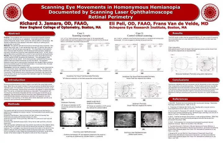
Scanning Eye Movements in Homonymous Hemianopia Documented by Scanning Laser Ophthalmoscope Retinal Perimetry. Richard J. Jamara, OD, FAAO, New England College of Optometry, Boston, MA. Eli Peli, OD, FAAO, Frans Van de Velde, MD Schepens Eye Research Institute, Boston, MA. Results.

E N D
Scanning Eye Movements in Homonymous Hemianopia Documented by Scanning Laser Ophthalmoscope Retinal Perimetry • Richard J. Jamara, OD, FAAO, • New England College of Optometry, Boston, MA Eli Peli, OD, FAAO, Frans Van de Velde, MD Schepens Eye Research Institute, Boston, MA Results Abstract Case 1: Scanning example Case2: Control without scanning Purpose.Young patients with homonymous hemianopia frequently develop scanning eye movements as an adaptation. Automated perimetry of these patients frequently shows ”vision” across the vertical midline in hemianopia. We wanted to determine if this apparent restoration might be an artifact due to scanning eye movements. Methods: Two patients with left homonymous hemianopia were examined. One patient was young, age 17 with hemianopia since age 12, and the other was an adult age 42, with hemianopia since age 40. The visual fields of both patients were measured with Humphrey 120 full field automated perimetry, Goldmann perimetry, and with the Scanning Laser Ophthalmoscope (SLO). In SLO, direct retinal observations permitted us to correct for eye movements during perimetry. Results.The young patient showed an extension of the visual field across the vertical midline on both Humphrey and Goldmann. The adult patient’s field respected the vertical midline. SLO retinal perimetry confirmed that the young patient’s field was indeed hemianopic as was the adult’s. The apparent extensions were due to scanning eye movements and variable fixation positions were documented as part of the SLO examination. In contrast to this, the adult patient demonstrated stable foveal fixation. Conclusion: Hemianopic adaptation with eye movements may be interpreted as vision restoration when measured by conventional perimetry. The SLO retinal perimetry may separate real visual restoration from the artifact caused by eye movements. Field extension due to scanning eye movements may improve the visual function just as much as actual field restoration might for homonymous hemianopia patients. The SLO results confirm that the expanded field for J.A. was a result of scanning eye movements and not a real restoration of the blind field. A similar case was reported by. Peli. Prism intervention: Both patients were fit with 40 diopter field expansion prisms and as shown here both patients experienced an expanded field. J.A., a 17 y.o. had a brainstem hemorrhage at age 12. He presented with clearly visible eye scanning enabling him to appear to see well into the left visual field on confrontation visual field measurements. A.A., a 42 y.o., suffered a recent stroke that resulted in a complete left homonymous hemianopia. A.A. did not appear to develop enhanced scanning. Case 2 Case 1 OS OD OS OD Binocular Goldmann Perimetry using prism intervention Humphrey Full Visual Field Automated Perimetry VF show an extension of the field across the vertical midline Humphrey Full Visual Field Automated PerimetryVisual Field loss respects the midline Introduction Conclusions Homonymoushemianopia is a loss of sight in one half of the visual field of both eyes. When this is the result of stroke or trauma sometimes the blind field may be restored in part. The restoration can be the result of spontaneous recovery, or an artifact of the perimetry. Scanning eye movements may appear to restore the blind field. This can be confused with spontaneous restoration. Young patients with homonymous hemianopia frequently develop scanning eye movements as an adaptation. We demonstrate that the Scanning Laser Ophthalmoscope (SLO) could differentiate between spontaneous restoration and apparent recovery secondary to eye scanning.In the SLO, direct retinal observation permits us to correct for eye movements during perimetry. Hemianopic adaptation with eye movements may be interpreted as vision restoration when measured by conventional perimetry. The SLO retinal perimetry may identify and separated real visual restoration from the artifact caused by eye movements. Field extension due to scanning eye movements may improve the visual function just as much as actual field restoration might for homonymous hemianopia patients. Methods for training in scanning eye movements may serve as a useful rehabilitation tool. References OS Goldmann Perimetry VF shows an extention of VF across the vertical the vertical midline OS OD BINOCULAR FIELDGoldmann Perimetry VF shows an extention of VF across the vertical the vertical midline Methods 1.Sabel BA. Residual vision and plasticity after visual system damage. Restorative Neurology and Neuroscience 1999; 15:73-79. 2. Timberlake GT, Mainster MA, Peli E, et al. Reading with a macular scotoma. Invest Ophthalmol Vis Sci 1986; 27:1137-1147. 3. Van de Velde FJ, Timberlake GT, Jalkh AE, Schepens CL. Static microperimetry with the laser scanning ophthalmoscope. Ophthalmologie 1990 May-June;4(3):291-4 (Article in French) -Translated by Amy Roan. 4. Peli E. Treating hemianopia using prisms to create peripheral diplopia. [Web Site] 1999; http://www.eri.harvard.edu/faculty/peli/papers/vision99/Hemianopia.html [Accessed 8 November 1999]. 5. Peli E. Field expansion for homonymous hemianopia by optically induced peripheral exotropia. Optom Vis Sci 2000; 77(9): 453-464. 6. Trauzettel-Klosinski S, Reinhard J. Macular Sparing and Fixation Behavior in Hemianopia. Vision Rehabilitation:Assessment,Intervention, and Outcomes/edited by Cynthia Stuen”Selected papers from Vision ’99: International Conference on Low Vision, July 1999. 7. Bergsma DP, van der Wildt GJ. Visual training of People with Visual Field Defects.Vision Rehabilitation:Assessment,Intervention, and Outcomes/edited by Cynthia Stuen”Selected papers from Vision ’99: International Conference on Low Vision, July 1999. Goldmann PerimetryVisual Field loss respects the midline Two patients with left homonymous hemianopia requested visual field testing to determine their eligibility for a driver’s license. The visual fields of both patients were tested using: Humphrey Field Analyzer: Static perimetry, full field 120 point Screening Test. Performed monocularly with standard stimulus III, white. Goldmann Perimeter::Kinetic perimetry. With white IV e stimulus for each eye and binocular field. Scanning Laser Ophthalmoscope (SLO): Using manual static perimetry using two target sizes. The scanning laser ophthalmoscope may present visual stimuli on the patient’s retina at light levels that are within normal environmental limits and safe for continuous exposure. The investigator may view and document the location of the stimuli on the patient’s retina as well as the patient’s fixation during the actual exam. The results can be recorded as a video image demonstrating both the perimetric results and the location of the fixation target on the patient’s retina at the time of response. OS OD OS OD Scanning Laser Ophthalmoscope Confirms hemianopic VF and apparent extensions the result of scanning as evidenced by variable fixation (+) positions Scanning Laser Ophthalmoscope Stable foveal fixation