Download
1 / 18
200 likes | 319 Views
Explore the structures and contents of the temporal and infratemporal fossae, including muscles of mastication, nerves, arteries, joints, and ligaments. Learn about the movements and relations of the temporomandibular joint.

E N D
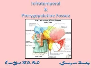
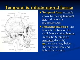
Temporal & infratemporal fossae • Temporal fossa :extends above by the sup.temporal line and below by zygomatic arch. • Infratemporal fossa : lies beneath the base of the skull, between the pharynx (medially) & ramus ofmandible (laterally). or the space lying below the temporal fossa and behind the maxilla.
Muscles of mastication:1-Temporalis • It lies in the temporal fossa. • Origin:floor of temporal fossa & temporal fascia. • Insertion:by a tendon into the coronoid process of the mandible. • N.supply: deep temporalnerves from the ant.division ofmandibular N. • Action: anterior fibers --- elevate the mandible.posterior fibers--- retract themandible.
Muscles of mastication • 2-Masseter muscle : • Origin :lower border & inner surface of zygomatic arch. • Insertion : lateral (outer) surface of ramus of the mandible. • N.supply :masseteric N. from anterior division ofmandibular N. • Action : raises the mandible.
Muscles of Mastication attached to mandible : Medial Surface Lateral Surface
Contents of the temporal fossa 1-Temporalis muscle. 2-Temporal fascia---- covers temporalis muscle, attached above to sup.temporal line and below to upper border of zygomatic arch. 3-Deep temporal nerves :from the ant. division ofmandibularN., emerge from upper border of lateral pterygoid, enter the deep surface of temporalis .
Contents of the temporal fossa 4-Auriculotemporal nerve : arise from the posterior division of mandibular N.It emerges from upper border of parotid gland ,It liesbehind superficial temporal artery & TMJ, in front of the auricle. It supplies skin of auricle , ext.auditory meatus and the scalpe over the temporal region.
Contents of the temporal fossa 5-Superficial temporalartery : it is a terminal branch of ext.carotid artery. • It Emerges from upper border of parotid gland, behind T.M.J. • It crosses root of zygomatic arch in front of auriculo-temporal N. & auricle ,here its pulsation can be easily felt.
Contents of Infratemporal fossa • Lateral & medial pterygoid muscles (muscles of mastication) • Branches of the mandibular N. • Otic ganglion. • Chorda tympani. • Maxillary artery. • Pterygoid venous plexus.
Lateral pterygoid • Origin :upper head---- from the infratemporal surface of the greater wing of sphenoid.Lower head---- from the lateral surface of lateral pterygoid plate. • Insertion :neck of mandible (pterygoid fovea) & articular disc of T.M.J. • N.supply :anterior division.of mandibular N. • Action:1-Pulls the neck of mandible forward with the articular disc to depress mandible during opening of mouth. 2-Acting with medial pterygoid of the same side during movement of chewing. 3-Acting with medial pterygoid to protrude the mandible.
Medial pterygoid • Origin:superficial head----- from the tuberosity of themaxilla.Deep head----- from the medial surface of the lateral pterygoid plate. • Insertion: angle of mandible (medial surface). • N.supply : main trunk ofmandibular N. • Action : 1-elevates the mandible. 2-Acting with lateral pterygoid duringmovement of chewing.
Tempromandibular joint (TMJ) • Articlation :between the articular tubercle & mandibular fossa of temporal bone, and the head of mandible (condyloid process). • Type :condyloid synovial joint. • Capsule :it surrounds the joint. • Synovial membrane--- lines the capsule in upper & lower cavities.
Ligaments of Temperomandibular joint : • Lateral temporomandibular ligament : lies on the lateral side of joint ,between the tubercle and lateral surface of the neck of mandible. • Sphenomandibular ligament : lies on the medial side of the joint ,it connects the spine ofsphenoid to the lingula of mandibular foramen. • Stylomandibular ligamentbehind& medial .to the joint. it is a band ofthickened deep cervical fascia, from apex of styloid process to angle of mandibule.
Intracapsular articular disc • It is a plate offibro-cartilage, it divides the joint into upper & lower cavities. • It is attached in front to the tendon of lat. pterygoid , and by fibrous bands to head ofmandible. • Its upper surfaceis concavo-convexto fit the articular tubercle & mandibular fossa , while its lower surface is concave to fit the head of mandible.
N.supply-auriculotemporal & masseteric branches ofmandibular N. Movements: • Depression of mandibule : by lat.pterygoid, helped bydigastric,geniohyoid & mylohyoid muscles. • Elevation :by temporalis,masseter, and medial pterygoid. • Protrusion : by lateral + medial pterygoids of both sides. • Retraction :by post.fibers of temporalis . • Lateral chewing movement:by lat.& med. Pterygoids of both sides acting alternately.
Relation of the Temporomandibularjoint (TMJ) : • Anteriorly :mandibular notch and masseteric N. & artery (structures passing through mandibular notch). • Posteriorly :ext.auditorymeatus, glenoid process of parotid gland., auriculotemporal N., & superficial temporal artery. • Laterally :parotid gland, fascia & skin. • Medially :maxillary vessels.
Clinical significance of the TMJ : • The great strength of the Lat.TM ligamentprevents head of mandible from passing backward to cause fracture of the tympanic plate in case of severe blow on the chin. • The articular disc may be partially detached causing noisy & audibleclick, during movements of the joint.
Dislocation of the TMJ • Sometimes occurs when themandible is depressed. • In case of minor blow on chin or sudden contraction of lateral pterygoids as in yawning, leads to pull the head of mandible & articular disc forward beyond the summit of tubercle. • Reduction of disloction : by pressing the thumbs downward on the lower molar teeth and pushing the jaw backward.